Synonyms
Keloidal blastomycosis or Lobo’s disease
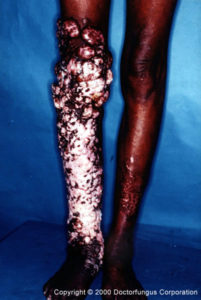
Lobomycosis of leg
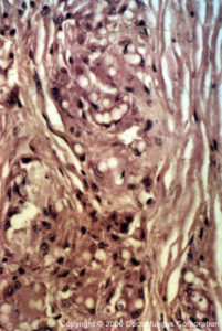
H & E stained tissue infected with Lacazia loboi
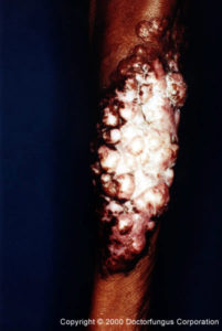
Arm with Lobomycosis
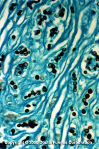
GMS stain of tissue infected with Lacazia loboi
Definition
Lobomycosis is a rare chronic cutaneous infection caused by N/A(L):Lacazia loboi previously called Loboa loboi. The disease manifests as keloids, verrucoid to nodular lesions, crusty plaques, and tumors. The fungus grows as globose cells that are connected to each other by a narrow neck. The cells may form branching chains. Developing lesions are well defined, smooth, painless and easily moved around since they lie free over the deeper tissues. Older lesions typically become verrucoid and ulcerative with satellite lesions resulting from autoinoculation [347, 1114, 1944].
<2>Clinical forms
Cutaneous and subcutaneous.
Prognosis and therapy
Lesions are managed by surgical excision. Since frequent relapse occurs, the excision must be wide. Surgery may result in new lesions [1943].
Histopathology
Nodules consist of subepidermal histiocytic granulomas that lie between the overlying skin and subcutaneous tissue. Fibrous tissue is dispersed between large numbers of giant cells and histiocytes. The giant cells are 40-80 µm in diameter. In older lesions, pyogenic infiltrates, parakeratosis, and acanthosis are present. Pseudoepitheliomatous hyperplasia and intraepidermal abscesses are absent. The fungus occurs as chains of globose cells 7-14 µm (average 9-10 µm) in diameter. Each cell is connected to the adjacent cell by a narrow neck. Some yeast cells occur within giant cells and macrophages, but the majority surround these cells.
Laboratory
Direct examination
Direct examination of clinical material, cutaneous and subcutaneous tissue, is mounted in 10% KOH and examined for the presence of chains of globose cells.
Isolation
Isolation of the fungus has never been achieved, except in mice foot pads.
Natural habitat
Unknown, but the infection is also found in dolphins