
Title:
Breakthrough Pulmonary Fusariosis During Therapeutic Posaconazole Prophylaxis in Acute Myeloid Leukemia
Submitted by: Taghrid Taha, MD, William Lainhart, PhD, D(ABMM), Saman Nematollahi, MD
Institution:
Banner Health-University of Arizona College of Medicine – Tucson
Email:
taghreedtarek111@gmail.com
Date Submitted:
May 2026
History:
A 66-year-old man with recently diagnosed acute myeloid leukemia (AML) transformed from high-risk hypoplastic myelodysplastic syndrome, presented with neutropenic fever during cycle 1 azacitidine/venetoclax therapy. His course had been complicated by severe pancytopenia requiring multiple transfusions. He was receiving posaconazole prophylaxis with documented therapeutic levels (>1 µg/mL).
He presented from the hematology clinic with fever (Tmax 38.4°C), gingival bleeding, fatigue, and generalized weakness. He denied cough, dyspnea, chest pain, gastrointestinal symptoms, urinary symptoms, or neurologic symptoms.
Relevant epidemiologic exposures included occupational exposure to construction and mechanical trades, and a 45-year tobacco use history. There were no recent travel, animal exposure, or sick contacts.
Physical Examination:
- Temperature: 38.4°C
- Heart rate: 102 bpm
- Blood pressure: 120/70 mm Hg
- Oxygen saturation maintained on room air.
The patient appeared chronically ill but in no acute distress. Pulmonary examination was clear bilaterally. No sinus tenderness was present. Skin examination demonstrated ecchymoses over the extremities without nodular or necrotic lesions. Neurologic examination was non-focal.
Laboratory Examination:
CBC:
- WBC: 0.6 ×10⁹/L
- ANC: 0.18 ×10⁹/L
- Hemoglobin: 7 g/dL
- Platelets: 10 ×10³/µL
Microbiology:
Positive:
- Respiratory pathogen panel: rhinovirus
Negative:
- Blood cultures
- MRSA nasal culture
- β-D-glucan (<31 pg/mL)
- Coccidioidomycosis serology/complement fixation
- Histoplasma urine antigen
- Legionella urine antigen
- Streptococcus pneumoniae urine antigen.
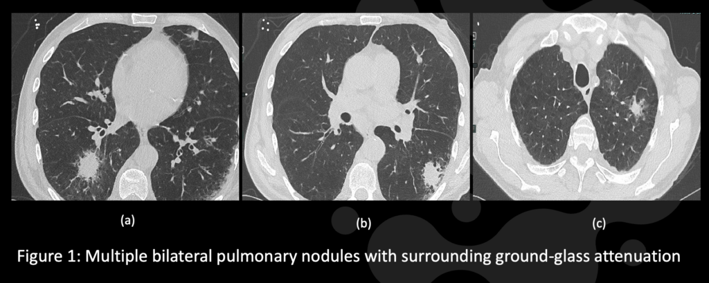
CT chest demonstrated:
- Multiple bilateral pulmonary nodules
- Surrounding ground-glass attenuation (“halo sign”)
- Mild mediastinal lymphadenopathy
Question 1: What are probable/possible diagnoses?
- Invasive pulmonary aspergillosis
- Pulmonary fusariosis
- Pulmonary mucormycosis (Rhizopus)
- Nocardiosis
- Mycobacterium avium complex infection
The patient was empirically started on vancomycin and cefepime for neutropenic fever. Vancomycin was discontinued after negative MRSA screening.
Persistent fevers despite broad-spectrum antibiotics and mold-active prophylaxis raised concern for breakthrough mold infection.
Microbiology/Diagnostic Tests Performed:
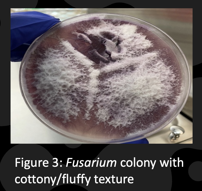
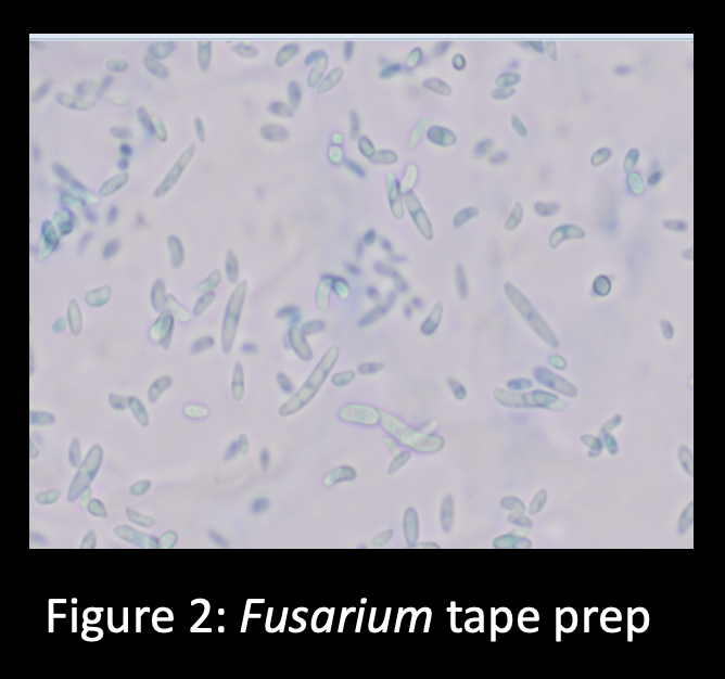
Fungal sputum cultures identified Fusarium species, demonstrating:
- Banana/crescent-shaped macroconidia
- Cottony/fluffy colonies on Sabouraud dextrose agar
Susceptibility testing demonstrated:
- Amphotericin B MIC: 1 µg/mL
- Voriconazole MIC: 2 µg/mL
- Posaconazole MIC: >16 µg/mL
- Isavuconazole MIC: >16 µg/mL
- Itraconazole MIC: >16 µg/mL
Final Diagnosis:
Pulmonary Fusariosis
Question 2: What treatment is recommended in the care of this patient?
Recommended treatment for invasive fusariosis includes:
- Voriconazole and/or liposomal amphotericin B (L-AMB)
- Consideration of combination antifungal therapy in severe disease
- Recovery of neutrophil counts/immunologic recovery
- Susceptibility-guided antifungal therapy
Emerging salvage agents include fosmanogepix and olorofim.
Treatment:
The patient was started on voriconazole 200 mg orally twice daily with planned therapeutic drug monitoring.
Combination therapy with liposomal amphotericin B was discussed but declined by the patient.
Repeat CT chest imaging was planned to assess treatment response. Dermatology follow-up was arranged because of concern for skin cancer risk associated with prolonged voriconazole therapy.
Outcome:
The patient remained clinically stable on room air and was discharged with outpatient follow-up in infectious diseases and hematology. Repeat chest imaging was planned to assess radiographic response to therapy.
Discussion:
This case highlights two major teaching points: (1) invasive fusariosis can occur as a breakthrough infection in profoundly neutropenic patients despite therapeutic posaconazole prophylaxis, and (2) the halo sign is not specific for Aspergillus and should prompt consideration of alternative angioinvasive molds such as Fusarium.
The patient had several important host risk factors predisposing to invasive mold infection, including AML on azacitidine/venetoclax therapy, profound, prolonged neutropenia, and environmental exposure through construction and mechanical work. Although invasive pulmonary aspergillosis was initially strongly considered because of the halo sign on CT imaging, persistent fevers despite mold-active azole prophylaxis raised concern for breakthrough infection with resistant molds such as Fusarium or Mucorales.
Invasive fusariosis is a severe opportunistic infection occurring predominantly in immunocompromised hosts, particularly patients with hematologic malignancies and prolonged neutropenia, with reported mortality frequently exceeding 50% (1). Contemporary leukemia cohorts demonstrate that most affected patients are neutropenic, with increasing incidence over time likely related to antifungal selective pressure. Importantly, breakthrough invasive fusariosis is increasingly recognized during mold-active azole prophylaxis (3). In this patient, susceptibility testing demonstrated high-level resistance to posaconazole, supporting breakthrough infection despite therapeutic drug levels.
Clinically, Fusarium most commonly presents with pulmonary disease but may disseminate hematogenously with fungemia and characteristic skin lesions (1,3). Although this patient did not demonstrate fungemia or cutaneous lesions, pulmonary nodules with surrounding ground-glass attenuation suggested angioinvasive fungal disease.
Radiographically, the halo sign reflects vascular invasion and hemorrhage but is not specific for aspergillosis, as similar findings occur with Fusarium and other molds (3,4). Therefore, microbiologic confirmation remains essential. Unlike Aspergillus, Fusarium species are more likely to grow from routine cultures and blood cultures during disseminated infection, potentially facilitating earlier diagnosis (1). In this case, fungal sputum culture identified Fusarium species by demonstrating characteristic banana/crescent-shaped macroconidia and cottony/fluffy colony morphology on Sabouraud dextrose agar.
Diagnosis remains challenging because non-culture fungal biomarkers have important limitations. Serum β-D-glucan lacks specificity, while galactomannan assay sensitivity is variable and species-dependent (2,5). Negative fungal biomarkers, therefore, do not exclude invasive fusariosis, particularly in neutropenic hosts.
Management of invasive fusariosis remains difficult because Fusarium species exhibit intrinsic multidrug resistance and highly variable susceptibility profiles (5,6). Current recommendations support voriconazole and/or liposomal amphotericin B, frequently in combination for severe disease (2). However, outcomes remain poor despite aggressive therapy. Emerging antifungal agents such as fosmanogepix and olorofim demonstrate promising in vitro activity against resistant Fusarium species and may represent future salvage options (6,7).
Combination salvage therapy incorporating terbinafine has been explored in severe invasive fusariosis, particularly in disseminated disease. In a small retrospective cohort (n=14), 93% of patients had disseminated cutaneous involvement and most received combination therapy with voriconazole plus liposomal amphotericin B. Despite partial skin response in 64% of patients, systemic progression occurred in 71%, with day-42 mortality also reaching 71%, highlighting the persistently poor outcomes associated with disseminated fusariosis despite aggressive combination antifungal therapy (8)
Ultimately, host immune recovery, particularly neutrophil recovery, remains the strongest predictor of survival and the most important determinant of outcome in invasive fusariosis (1,3).
Key References:
- Nucci M, Anaissie E. Fusarium infections in immunocompromised patients. Clin Microbiol Rev. 2007;20(4):695–704. PMID: 17934079.
- Tortorano AM, Richardson M, Roilides E, et al. ESCMID/ECMM guidelines for hyalohyphomycosis. Clin Microbiol Infect. 2014;20(Suppl 3):27–46. PMID: 24548001.
- Matsuo T, Wurster S, Jiang Y, et al. Invasive fusariosis in patients with leukemia in the era of mould-active azoles: increasing incidence, frequent breakthrough infections and lack of improved outcomes. J Antimicrob Chemother. 2024;79:297–306. PMID: 38073151.
- Patterson TF, Thompson GR 3rd, Denning DW, et al. Practice guidelines for aspergillosis. Clin Infect Dis. 2016;63(4):e1–e60. PMID: 27365388.
- Wiederhold NP. Antifungal resistance: current trends. Clin Infect Dis. 2017;64(Suppl 2):S130–S134. PMID: 28475798.
- Al-Obaidi MM, Nematollahi S, Hayes J, Nix DE. Past, present, and emerging antifungals against Fusarium and other rare non-Aspergillus hyalohyphomycetes: a narrative review. Expert Opin Pharmacother. 2026. PMID: 41877374.
- Berkow EL, Lockhart SR. Activity of fosmanogepix against molds. Antimicrob Agents Chemother. 2018;62(11):e01120-18. PMID: 30104208.
- . Matsuo T, et al. Combination therapy with terbinafine for invasive fusariosis. Open Forum Infect Dis. 2024.