(described by Viala and Bayer in 1891)
Berkhout, 1923 nom. cons.
Taxonomic Classification
Kingdom: Fungi
Phylum: Ascomycota
Class: Euascomycetes
Order: Dothideales
Family: Dothioraceae
Genus: Aureobasidium (Teleomorph of Aureobasidium pullulans is Discosphaerina fulvida)
Description and Natural Habitats
Aureobasidium is a cosmopolitan, dematiaceous fungus commonly isolated from plant debris, soil, wood, textiles, and indoor air environment.
Species
The genus Aureobasidium includes 14 species and one variety. Among these, Aureobasidium pullulans is the only well-known species. The discussion on this page in general includes only Aureobasidium pullulans. See the list of obsolete names and synonyms for older names of these species.
Pathogenicity and Clinical Significance
Aureobasidium pullulans is one of the causative agents of phaehyphomycosis. It may cause keratomycosis, pulmonary mycosis with sepsis and other opportunistic infections, as well as cutaneous mycoses such as eumycotic dermatitis. While Aureobasidium mansoni was reported to cause nosocomial meningitis [1089], Aureobasidium pullulans was isolated in patients with peritonitis [1847]. Aureobasidium may also colonize hair, skin, and nails in humans. The pathogenicity of Aureobasidium remains limited and uncommon [1847]. Thus, Aureobasidium pullulans is commonly considered as a contaminant.
Macroscopic Features [462, 1295, 2144]
It grows moderately rapidly and matures within 7 days of incubation. The colony diameter is 1 to 3 cm following incubation at 25°C for 7 days on potato glucose agar. The colonies are flat, smooth, moist, yeast-like, mucoid to pasty, shiny and leathery in appearance. The surface is white, pale pink or yellow at the beginning and becomes brown to black and velvety with a grayish fringe by aging. Reverse is pale or black.
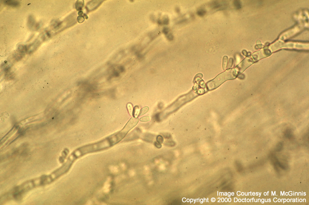
Microscopic Features [462, 1295, 2144]
When the colony is young, moist, and yeast-like, unicellular budding yeast cells (blastoconidia) are the only structures observed microscopically. By aging, while the colony gets black and velvety, hyphae become visible.
Blastoconidia are pale in color. The synchronous development of blastoconidia in tufts is typical. Hyphae are septate. They appear hyaline at the beginning and get dark brown by aging. The width of the hyphae is generally 2-10 µm but may be as thick as 15-20 µm. Aureobasidium has no distinct conidiophores. Conidiogenous cells, which are not much differentiated, are either intercalary or located terminally in the hyphae. The conidia (4-6 x 2-3 µm in size) are one-celled, hyaline and oval to cylindrical in shape. They form clusters or are located along the hyphae. These conidia continue to multiply by budding and form secondary blastoconidia.
As well as blastoconidia, chlamydoconidia and arthroconidia may also be observed. The hyphae of some strains differentiate to form thick-walled, large (up to 12×6 µm in size), phaeoid (brown in color) chlamydoconidia. Thick-walled, one- to two-celled, phaeoid arthroconidia are produced in old, mature cultures.
Histopathologic Features
As for other agents of phaehyphomycosis, dark-walled, short, septate hyphae may be observed in H&E stained sections. If the pigment production is not prominent, the use of Fontana-Masson silver stain may be advantageous [462].
Compare to
Hormonema dematioides, Phaeococcomyces
Key Features for Differentiation from Similar Genera [1295, 2144]
| STRUCTURE/PROPERTY | DIFFERENTIATION |
|---|---|
| Blastoconidia formation: synchronous and in close tufts | Aureobasidium (+) Hormonema* (-) |
| Hyaline blastoconidia | Aurebasidium (+) Phaeococcomyces (-) |
* produces blastoconidia successively, in a chain-like fashion and from a single opening
Laboratory Precautions
No special precautions other than general laboratory precautions are required.
Susceptibility
No susceptibility data are available for Aureobasidium. In vitro susceptibility testing methods are not yet standardized for dematiceaous fungi.