(described by Eidam in 1886)
Taxonomic Classification
Kingdom: Fungi
Phylum: Zygomycota
Subphylum: Zygomycotina
Class: Zygomycetes
Order: Entomophthorales
Family: Basidiobolaceae
Genus: Basidiobolus
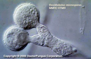
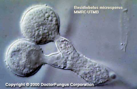
Globose spores which have not been discharged from the sporophore of Basidiobolus microsporus
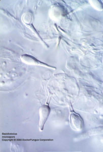
Elongated spore on a sporophore of Basidiobolus ranarum
Description and Natural Habitats
Basidiobolus is a filamentous fungus isolated from dung of amphibians, reptiles, and insectivorous bats, as well as wood lice, plant debris and soil. Although it is cosmopolitan, the human infections due to Basidiobolus are reported mostly from Africa, South America, and tropical Asia.
Species
Although the pathogenic Basidiobolus isolates have once been identified as separate species, as Basidiobolus ranarum, Basidiobolus meristosporus, and Basidiobolus haptosporus, recent work on their antigens, restriction analysis of rDNA, and isoenzyme banding show that all Basidiobolus isolates that are pathogenic to humans belong to a unique species, Basidiobolus ranarum. See the list of obsolete names and synonyms for older names of these species.
Pathogenicity and Clinical Significance
Basidiobolus ranarum is the etiologic agent of subcutaneous chronic zygomycosis in man.
This infection is also called entomophthoromycosis basidiobolae. It is characterized by its granulomatous nature and formation of hard, nonulcerating subcutaneous masses at limbs, chest, back, and buttocks. Systemic infection is rare. Basidiobolus is a true pathogen, causing infections in immunocompetent host. However, recent data on angioinvasive infections due to Basidiobolus in immunocompromised patients suggest that it is emerging as an opportunistic pathogen as well [1918]. It may rarely cause acute zygomycosis, which is similar to that defined for the zygomycetous fungi in the Mucorales order. Gastrointestinal infections due to Basidiobolus ranarum have also been reported in man [2490].
Basidiobolus can cause infections in animals as well as humans. Cutaneous lesions in amphibians, subcutaneous infection in horses and gastrointestinal lesions in dogs have so far been reported.
Macroscopic features
It grows moderately rapidly, the diameter of the colony reaching 1 to 3 cm following incubation at 25°C for 7 days on potato glucose agar. Its growth is more rapid at 30°C compared to 37°C. The colony is flat, thin, waxy and buff to gray in color initially. It later becomes heaped up or radially folded, grayish brown in color and is covered by fine, white, powdery surface mycelium. The reverse is white or pale. The germinated conidia ejected from the primary colony may form satellite colonies. Some Basidiobolus strains have an earthy odor typical for Streptomyces spp. [462, 1295, 2144].
Microscopic Features [462, 1295, 2144]
Sporulation is observed only in freshly isolated strains and ceases as the colony gets older or is subcultured. Thus, the strain should be examined microscopically as soon as it is isolated. Special medium containing glucosamine hydrochloride and casein hydrolysate may be used to stimulate sporulation.
The hyphae are large (8 to 20 µm in diameter) and septate. The number of septa increases and becomes plenty as the spores are produced. The sexual spores, zygospores of Basidiobolus, are thick-walled and smooth or have undulating outer cell walls. Each zygospore typically possesses two lateral appendages which are the conjugation beaks. These appendages are the remnants of a copulatory tube.
The sporangiophores (sporophores) of Basidiobolus are of two types and can produce two different types of asexual conidia, the sporangioles. The first type of sporangiophore which has a subterminal swelling, produces and discharges ejectable, globose to pyriform unispored primary sporangioles from its apex. This single cell spore also carries a fragment from the sporangiophore. This is primarily due to the tearing of the sporangiophore wall due to the internal pressure during ejection. These sporangioles are also called ballistospores. The second type of sporangiophores which are not swollen but are narrow, passively liberate unispored, clavate-shaped secondary sporangioles. The apex of this passively released spore has a knob-like adhesive tip. These spores may also function as sporangia and produce several sporangiospores.
Histopathologic Features
In tissue sections infected with Basidiobolus ranarum, broad, thin-walled hyphae which are aseptate or infrequently septate are observed. The contours of the hyphae are nonparallel and branches are random.
In general, in contrast to those in Mucorales order, fungi included in Entomophthorales order do not invade vascular tissue. Thus, no infarction is observed in infected tissue. However, Basidiobolus ranarum infections may be angioinvasive in immunocompromised hosts [1918]. There is a chronic inflammatory response with a predominance of eosinophiles [462].
Compare to
The macroscopic morphology is similar to that of Phytium spp.
Key Features for Differentiation
| STRUCTURE | DIFFERENTIATION |
|---|---|
| Ejected sporangioles possessing papilla | Basidiobolus (-) Conidiobolus (+) |
| Sporangiophores with swollen apices | Basidiobolus (+) Conidiobolus (-) |
| Zygospores | Basidiobolus (+) Conidiobolus coronatus (-) Conidiobolus incongruus (+) |
| Zygospores with conjugation beaks | Basidiobolus (+) Conidiobolus incongruus (-) |
Laboratory Precautions
No special precautions other than general laboratory precautions are required.
Limited data obtained so far suggest that, while flucytosine MICs are very high, those of fluconazole, itraconazole, ketoconazole, and miconazole are considerably low for Basidiobolus strains [912].
Infections due to Basidiobolus are difficult to treat. Systemic potassium iodide solution and trimethoprim-sulfamethoxazole are usually preferred. Oral ketoconazole and fluconazole may be of help in some cases [1895]. However, amphotericin B has practically no significant efficacy and surgery is not curative .