(described by Link ex Gray in 1821)
Taxonomic Classification
Kingdom: Fungi
Phylum: Ascomycota
Class: Euascomycetes
Order: Chaetothyriales
Family: Herpotrichiellaceae
Genus: Fonsecaea
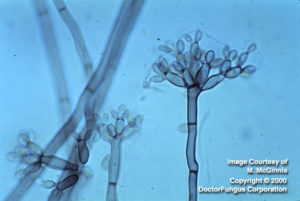
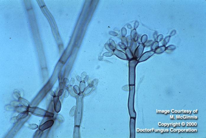
The primary conidia function as sympodial conidiogenous cells to form the secondary conidia
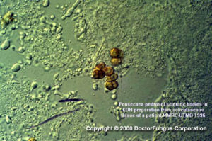
Muriform cells (Medlar bodies) from a case of chromoblastomycosis
Description and Natural Habitats
Fonsecaea is a pigmented (dematiaceous), filamentous fungus found in rotten wood and soil. It has no known teleomorphic phase. As well as being a saprophyte in nature, it causes infections in humans. Cold-blooded animals living in swamps may also be infected. Fonsecaea exhibits in vivo dimorphism; it produces a specific structure (sclerotic body) only in tissue and grows in mould form in laboratory conditions [462, 531, 1295, 2144, 2202].
Species
The genus Fonsecaea contains two species: Fonsecaea compacta and Fonsecaea pedrosoi.
Synonyms
See the summary of synonyms for Fonsecaea spp.
Pathogenicity and Clinical Significance
Fonsecaea is one of the causative agents of the post-traumatic, chronic infection of subcutaneous tissues known as chromoblastomycosis. Chromoblastomycosis presents with papules and verrucose cauliflower-like lesions most commonly on lower extremities [286, 462]. Primary nasal chromoblastomycosis has also been reported [2488]. The etiologic agents of chromoblastomycosis are generally members of three genera of dematiaceous fungi that inhabit the soil: Fonsecaea, Phialophora, and Cladosporium [533].
Fonsecaea pedrosoi is one of the major causative agents of chromoblastomycosis in humid tropical areas, specifically South America and Japan. Fonsecaea compacta, on the other hand, is a rare cause of chromoblastomycosis in tropical Central and North America [531, 2202]. Systemic invasion following chromoblastomycosis is very rare.
In addition to chromoblastomycosis, Fonsecaea may cause other human infections as well. Paranasal sinusitis [1506], keratitis [737], and fatal brain abscesses following hematogenous dissemination [784] have been reported.
Macroscopic Features
Fonsecaea grows slowly and produces restricted, flat to raised and folded, velvety to cottony colonies on potato dextrose agar at 25°C. The colonies mature in 14 days. From both front and reverse, they are olivaceous to brown-black in color. The filamentous appearance is maintained upon cultivation at 25, 30, or 37°C [531, 1295, 2202].
Microscopic Features
Fonsecaea produces septate, dark brown hyphae and suberect conidiophores that highly branch at apices. The conidiophores are pale brown, erect, septate, and sympodial with conidiogenous zones confined to the upper portion. The conidia (1.5-3 x 2.5-6 µm) are brown and barrel-shaped. Four types of conidiogenesis are observed [1295]:
- Fonsecaea type: Conidia are 1-celled and arise upon swollen denticles that are located at the tips of the conidiophores. These primary conidia function as sympodial conidiogenous cells, becoming irregularly swollen at their apices. These in turn give rise to 1-celled, pale brown, secondary conidia on swollen denticles. The secondary conidia often produce tertiary series of conidia like those formed by the primary conidia, resulting in a complex conidial head. Long chains of conidia are not formed in this type of conidiogenesis. This type of conidiogenesis is primarily observed for the strains belonging to the genus Fonsecaea.
- Cladosporium type: Conidiophores give rise to primary shield-shaped conidia which finally produce long, branching chains of oval, dematiaceous conidia. The conidia have visible dark hila which are actually the attachment scars. This type of conidiogenesis is primarily observed for the strains belonging to the genus Cladosporium, but may also be observed in strains of Fonsecaea.
- Phialophora type: In this type of conidiogenesis, conidia are located at the apices of the phialides which are vase-shaped and have collarettes. This type of conidiogenesis is primarily observed for the strains belonging to the genus Phialophora, but may also very rarely be observed in strains of Fonsecaea.
- Rhinocladiella type: Conidiophores are sympodial and have denticles which bear 1-celled, pale brown conidia. The conidia may be located at the tips and along the sides of conidiophores. Formation of secondary conidia is very rare. This type of conidiogenesis is primarily observed for the strains belonging to the genus Rhinocladiella, but may also be observed in strains of Fonsecaea.
Histopathologic Features
All fungi causing chromoblastomycosis produce a distinctive structure in infected tissues that is called a sclerotic body (sometimes one also sees the phrases muriform cell and Medlar body). Phaeoid (dark-colored) hyphae may also be observed in infected tissues. Sclerotic bodies are dark brown, spherical or polyhedral, thick-walled structures which have horizontal and vertical septa inside. They may be found singly, in clusters, or within the giant cells. Melanin, which is produced in the cell wall of the sclerotic body, gives the dark brown color to the structure. Unlike yeasts, sclerotic bodies mostly divide by separation along the septa. Budding may also occur but is not believed to be a significant means for reproduction. The production of sclerotic bodies only in tissue is believed to be regulated by the concentration of calcium ions within the infected tissue [462, 2482].
The presence of sclerotic bodies in the tissue confirms the diagnosis of chromoblastomycosis. The determination of the infecting fungus requires cultivation.
See also our histopathology page.
Compare to
Cladosporium
Cladophialophora
Rhinocladiella
Fonsecaea is recognized by its complex conidial head consisting of apically, irregularly swollen series of conidia that function as conidiogenous cells. This predominant type of conidial development distinguishes Fonsecaea from Cladosporium and Rhinocladiella, as described in the section on microscopic appearance. Fonsecaea is differentiated from Cladophialophora by producing short conidial chains which consist of 5 or less conidia.
Fonsecaea compacta differs from Fonsaceae pedrosoi by having subglobose to globose conidia occurring in compact heads, in contrast to the loose heads consisting of elongated conidia in Fonsaceae pedrosoi.
Fonsecaea should be handled with care in a biological safety cabinet.
Susceptibility
Few data are available and there is as yet no standard method for in vitro susceptibility testing of Fonsecaea spp. Amphotericin B, ketoconazole, miconazole, itraconazole, voriconazole, and terbinafine generate low MICs for Fonsecaea isolates. Itraconazole and voriconazole appear to have better in vitro activity than amphotericin B. Caspofungin is also active in vitro against Fonsaceae pedrosoi [558].Fluconazole, on the other hand, has no practical in vitro activity against Fonsecaea [1130, 1131, 1490, 1491, 1864].
For MICs of various antifungal drugs for Fonsecaea, see our N/A(L):susceptibility database.
Cryosurgery and itraconazole are currently used to treat cases of chromoblastomycosis [734, 1857, 1895]. Cryosurgery is preferred for small lesions, while itraconazole is used for larger lesions. The combination of the two therapeutic modalities may also be applied [286, 287]. Notably, terbinafine also appears promising in treatment of chromoblastomycosis [692]. However, chromoblastomycosis is difficult to treat and most therapeutic means provide only a modest success rate.