(described by Gruby in 1843)
Taxonomic classification
Kingdom: Fungi
Phylum: Ascomycota
Order: Onygenales
Family: Arthrodermataceae
Genus: Arthroderma (includes Nannizia, Microsporum, Epidermophyton, Trichophyton)
Description and Natural Habitats
Microsporum is a filamentous keratinophilic fungus included in the group of dermatophytes. While the natural habitat of some of the Microsporum spp. is soil (the geophilic species), others primarily affect various animals (the zoophilic species) or human (the anthropophilic species). Some species are isolated from both soil and animals (geophilic and zoophilic). Most of the Microsporum spp. are widely distributed in the world while some have restricted geographic distribution. Microsporum is the asexual state of the fungus and the telemorph phase is referred to as genus Arthroderma [358, 1796, 2144].
Species
The genus Microsporum includes 17 conventional species. Among these, the most significant are:
M. audouinii
M. gallinae
M. ferrugineum
M. distortum
M. nanum
M. canis
M. gypseum
M. cookei
M. vanbreuseghemii
| SPECIES | CLASSIFICATION (NATURAL RESERVOIR) |
|---|---|
| Microsporum audouinii | Anthropophilic |
| Microsporum canis | Zoophilic (Cats and dogs) |
| Microsporum cooeki | Geophilic (also isolated from furs of cats, dogs, and rodents) |
| Microsporum ferrugineum | Anthropophilic |
| Microsporum gallinae | Zoophilic (fowl) |
| Microsporum gypseum | Geophilic (also isolated from fur of rodents) |
| Microsporum nanum | Geophilic and zoophilic (swine) |
| Microsporum persicolor | Zoophilic (vole and field mouse) |
Molecular studies for taxonomic reclassification of Microsporum spp. are in progress [885].
The characteristics of Microsporum spp. are discussed in general in this page and the specific characteristics of each species are summarized in the Table following the sections about macroscopic and microscopic features. For detailed information about each species, please refer to the related page.
See the summary of synonyms and teleomorph-anamorph relations for the Microsporum spp.
Pathogenicity and Clinical Significance
Microsporum is one of the three genera that cause dermatophytosis. Dermatophytosis is a general term used to define the infection in hair, skin or nails due to any dermatophyte species. Similar to other dermatophytes, Microsporum has the ability to degrade keratin and thus can reside on skin and its appandages and remains noninvasive. As well as the keratinase enzyme, proteinases and elastases of the fungus may act as virulence factors. Notably, Microsporum spp. mostly infect the hair and skin, except for Microsporum persicolor which does not infect hair. Nail infections are very rare. The pathogenesis of the infection depends on the natural reservoir of the species. Geophilic spp. are acquired via contact with soil. Zoophilic species are transmitted from the infected animal. Direct or indirect (via fomites) human-to-human transmission is of concern for anthropophilic species. Asymptomatic carriage may be observed. As well as the otherwise healthy hosts, immunocompromised patients are also infected [54, 462, 657, 774, 806, 1192, 1679, 1959, 2400].
Macroscopic Features
Microsporum colonies are glabrous, downy, wooly or powdery. The growth on Sabouraud dextrose agar at 25°C may be slow or rapid and the diameter of the colony varies between 1 to 9 cm after 7 days of incubation. The color of the colony varies depending on the species. It may be white to beige or yellow to cinnamon. From the reverse, it may be yellow to red-brown. As are discussed on separate pages for each species, varieties in macroscopic morphology and color help in inter-species differentiation [2144].
Hair perforation test, ability to grow on rice grains and also at 37°C provide useful hints to differentiate Microsporum spp. from eachother.
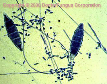
Microscopic Features
Microsporum spp. produce septate hyphae, microaleurioconidia, and macroaleurioconidia. Conidiophores are hyphae-like. Microaleuriconidia are unicellular, solitary, oval to clavate in shape, smooth, hyaline and thin-walled. Macroaleuriconidia are hyaline, echinulate to roughened, thin- to thick-walled, typically fusiform (spindle in shape) and multicellular (2-15 cells). They often have an annular frill. Inoculation on specific media, such as potato dextrose agar or Sabouraud dextrose agar supplemented with 3 to 5% sodium chloride may be required to stimulate macroconidia production of some strains. As are discussed on separate pages for each species, varieties in shape of macroconidia and abundance of microconidia help in inter-species differentiation [2144].
| Key to the More Common Species of Microsporum | |
|---|---|
| 1. | In vitro hair perforation test negative. (2) |
| 1.’ | In vitro hair perforation test positive. (4) |
| 2. | Diffusible strawberry-red pigment produced; macroconidia clavate to cigar shaped, with walls that are smooth to finely echinulate. M. gallinae |
| 3. | Colonies slow growing with many folds, glabrous to membranous, sometimes velvety, yellow to deep rust colored with a yellow to dull orange reverse; some hyphae often thick walled and highly segmented; usually non-sporulating, but forming fusiform macroconidia on rice grains or dilute Sabouraud glucose agar M. ferrugineum |
| 3.’ | Colonies slow growing, glabrous to velvety, whitish tan to brownish, reverse usually salmon in color; terminal vesicles with solitary spinelike projections are often present. M. audouinii |
| 4. | Macroconidia irregular, contorted. M. distortum |
| 4.’ | Macroconidia not contorted. (5) |
| 5. | Macroconidia typically 2- to 3-celled, obovoid, thin walled and rough. M. nanum |
| 5.’ | Macroconidia more than 3- celled, spindle-like or elliptic in shape. (6) |
| 6. | Colonies deep yellow or buff to cinnamon brown. (7) |
| 6.’ | Colonies white, yellowish tan, pale rose, or deep rose. (8) |
| 7. | Macroconidia spindle shaped, often with a predominant apical knob, with outer cell walls thicker than septal walls; colonies typically have a white to deep yellow color with a yellow to orange reverse. M. canis |
| 7.’ | Macroconidia elliptic with thin outer cell and septal walls, outer cell walls tend to collapse slightly between the septa; distinct annular frill present; colonies granular to powdery, buff to cinnamon brown. M. gypseum |
| 8. | Colonies yellowish, greenish buff to dark brown with red reverse color; macroconidia thick walled, 6- to 10- celled, 12-28 x 31-75 um. M. cookei |
| 8.’ | Colonies white to yellowish, tan or pink to deep rose, light yellow reverse color; macroconidia 5- to 12-celled, 8-13 x 44-88 um, cylindrical to cigar shaped. M. vanbreuseghemii |
Histopathologic Features
See our histopathology page.
Compare to
Microsporum differs from Trichophyton and Epidermophyton by having spindle-shaped macroconidia with echinulate to rough walls [2144].
Laboratory Precautions
No special precautions other than general laboratory precautions are required.
Susceptibility
In vitro susceptibility tests for dermatophytes are not yet standardized and available data are limited. Most investigators prefer to use the modifications of the NCCLS (M38P) methodology in the interim for testing the dermatophytes [1622]. Terbinafine and itraconazole appear active in vitro against Microsporum spp. However, this fungus was found to be the least terbinafine-susceptible dermatophyte in vitro in one of the studies [1119, 1554, 2282].
While griseofulvin was once the drug of choice for treatment of infections due to Microsporum as well as other dermatophytes, safer and more efficacious alternatives are now available and preferred. Oral therapy with terbinafine and itraconazole are very widely used for treatment of Microsporum infections [552, 657, 775, 1012, 1334, 1335].
For MICs of various antifungal drugs so far reported for Microsporum, see our susceptibility database.