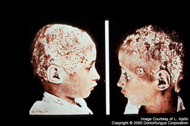
Tinea capitis and (A):Tinea favosa
Tinea capitis
Synonyms
Ringworm of the scalp and hair
Definition
Tinea capitis is the fungal infection of the scalp due to dermatophytes. The hair of head, eyebrows, eyelashes may also be involved. The skin may be involved alone, as is seen with infection due to N/A(L):Trichophyton rubrum.
There are three forms of hair infection. In the form known as endothrix, the infection begins by penetration of the hair, and the organism then grows up the interior main axis of the hair where it fragments into arthroconidia. A typical causative agent is T. tonsurans. The exception to the rule about arthroconidia formation is Trichophyton schoenleinii. This organism causes an endothrix-style growth, but without the arthrocondia. Instead, channels are formed within the hair shaft. This is useful diagnostically, as air bubbles move along these channels when an infected hair is immersed in a liquid. This later form of infection, as well as the clinical presentation of scutula, carries the special label (A):Tinea favosa, or favus [402, 657].
In the form of hair infection known as ectothrix, the infection begins as in endothrix, but it then extends back out through the hair cuticle (the outer wall of the hair) and forms a mass of arthroconidia both within and around the hair shaft. A typical causative agent is N/A(L):Microsporum canis [840].
Epidemiology
Tinea capitis is characteristically a fungal infection affecting children between 4 and 14 years of age [321]. Although a (A):large list of fungal pathogens have been implicated, Trichophyton tonsurans is responsible for more than 90% of cases in North America and the United Kingdom. Urban cases are commonly acquired from classmates or family members. Overcrowding, poor hygiene and protein malnutrition favor the occurrence of this disease [1447]. Cases caused by N/A(L):Microsporum canis occur sporadically and are acquired from puppies and kittens [1045].
Clinical manifestations
The diagnosis of tinea capitis should be suspected in any child older than 3 months with a scaly scalp. Differential diagnosis includes seborrheic dermatitis, atopic dermatitis, psoriasis, alopecia areata, trichotillomania, bacterial folliculitis, abscesses and neoplasias.
The forms of Tinea Capitis are:
| CATEGORIES | CLINICAL APPEARANCE |
|---|---|
| NonInflammatory Diffuse scaly Grey patch |
Erythematous papule(s) around the hair shaft appear initially. Subsequently, one or several patches of scaly alopecia are seen where the hairs are broken just above the level of the scalp. The hair looks lusterless and gray because it is covered with arthrospores. |
| Inflammatory Diffuse pustular Kerion |
Scattered painful pruritic pustular folliculitis generally associated with regional lymphadenopathy and even fever. In about 2-3 percent, boggy nodules studded with broken hairs and purulent sticky material (“kerion”) appear. Scarring alopecia develops subsequently. |
| “Black dot” | As it is related to the endothrix variety of infection, hairs become notably fragile and break easily at the level of the scalp. The rest of the infected follicle looks like “black dots”. Variable degrees of scaling and inflammation are seen. |
Prognosis and therapy
In contrast with other forms of dermatophytoses, tinea capitis should be treated with oral agents. Griseofulvin and ketoconazole have been extensively used, however terbinafine, itraconazole, and fluconazole are also recomended [616].
| DRUG | REGIMEN | NOTES |
|---|---|---|
| Griseofulvin | 10 mg/kg/day x 8-10 weeks | Ingest with fatty food to increase absorption |
| Itraconazole | 5 mg/kg/day x 1-4 weeks | Take oral solution without food |
| Terbinafine | < 20 kg: 62.5 mg/day 20 — 40 kg: 125 mg/day x 4 wks > 40 kg: 250 mg/day |
From [1045]
A pruritic papular rash that commonly affects the outer helix of the ears may appear after the introduction of therapy and should not be confused with a drug reaction [1045].
Some patients may need either intralesional or short oral courses of corticosteroids. Surgical approaches such as incision and drainage of kerion are not suggested. Bacterial superinfections are common, and antibiotic treatments may be neccesary [616, 1045].
Topical therapy with selenium sulphide, and povidone iodine shampoos are recommended twice a week to reduce infectivity, but should not be used as the sole therapeutic agents [1045].
Mycology(principal dermatophytes)
- Microsporum audouinii
- N/A(L):Microsporum canis
- N/A(L):Microsporum distortum
- N/A(L):Microsporum gypseum
- Trichophyton megninii
- Trichophyton mentagrophytes
- Trichophyton rubrum
- Trichophyton schoenleinii
- Trichophyton tonsurans
- Trichophyton verrucosum
Natural habitat
Animals, humans, soil
Tinea favosa
Synonyms
Favus
Definition
Tinea favosa is usually considered a variety of (A):Tinea capitis because it classically involves the scalp, however, this mycotic infection may also involve glabrous skin and nails.
Epidemiology
Favus is a disease of rural areas where poor hygiene and malnutrition are frequent [1447]. It generally affects groups of family, reason why it is postulated that prolonged and intimate contact is neccesary for transmission [1137].
Clinical manifestations
Favus is characterized by the occurrence of dense masses of mycelium and epithelial debris forming yellowish cup-shaped crusts called scutula. The scutulum develops at the surface of a hair follicle with the shaft in the center of the raised lesion. Removal of these crusts reveals an oozing, moist, red base. After a period of years, atrophy of the skin occurs leaving a cicatricial alopecia and scarring. Scutula may form on the scalp or the glabrous skin.
Prognosis and therapy
Therapeutic considerations for tinea favus are the same as for (A):Tinea capitis [616].
Mycology
- N/A(L):Microsporum gypseum
- Trichophyton schoenleinii
Natural habitat
Humans, soil