Title: Penile Lesions as Initial Manifestation of Invasive Fusariosis
Submitted by: Apoorva Dharmadhikari MD, Dimitrios Kontoyiannis MD, PhD
Institution: University of Texas MD Anderson Cancer Center
Email: ARDharmadhikari@mdanderson.org
Date Submitted: 3/25/26
History:
A 72-year-old man with a past medical history of chronic lymphocytic leukemia (CLL), chronic sinusitis with prior sinus surgery, was hospitalized for 2 months in El Paso, Texas, for a new diagnosis of high-risk myelodysplastic syndrome (MDS) with excess blasts. He received two cycles of chemotherapy with decitabine and venetoclax without improvement in his cell counts. During this time, he had been receiving prophylactic oral acyclovir and fluconazole for prolonged neutropenia. He was subsequently transferred to MD Anderson Cancer Center for consideration of stem cell transplant for definitive treatment.
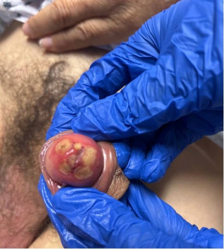
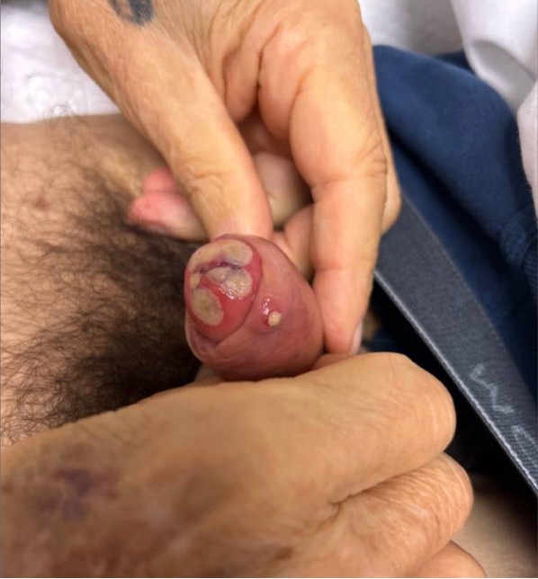
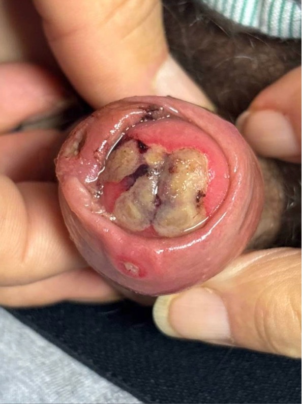
On admission to MD Anderson, the patient was noted to have neutropenic fever with a temperature of 100.6°F and a white blood cell count of 0.1 ×10⁹/L. When asked about new symptoms, he reported painful penile lesions that had first appeared one month prior and had progressively worsened over the last two weeks. The lesions had become ulcerated and increasingly painful, causing difficulty with voiding. He also endorsed mild nasal congestion.
Given the unusual genital lesions, Infectious Disease was consulted.
Social History: The patient was originally from Mexico but had lived in El Paso, Texas, for many years, working as a carpenter and woodworker. His hobbies included gardening and using his jacuzzi. He denied recent international travel, but had recently gone camping in Ruidoso, New Mexico (approximately 6,000 feet elevation). He denied animal exposures and high-risk sexual behaviors.
Physical Examination:
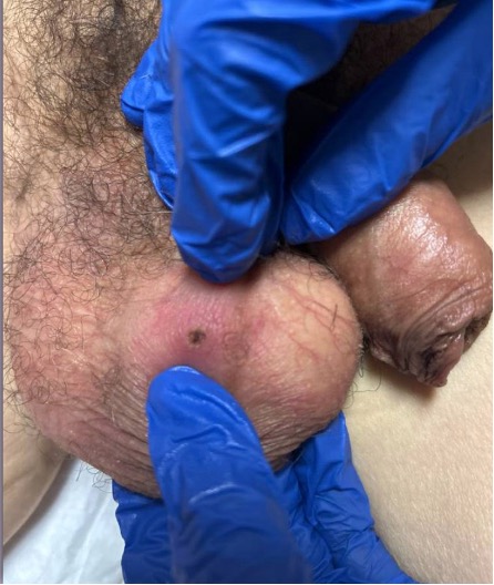
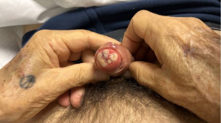
Dermatologic examination revealed multiple round, well-demarcated friable ulcerations involving the glans penis, foreskin, and scrotum. The lesions were tender with surrounding erythema but without purulent drainage.
No inguinal lymphadenopathy was present. No testicular masses were palpated.
ENT evaluation with nasal endoscopy demonstrated bilateral diffuse crusting of the nares with fungal-appearing debris and dark crusting along the nasal septum extending superiorly.
The remainder of the physical examination was unremarkable.
Laboratory Examination:
Laboratory studies demonstrated profound pancytopenia:
- White blood cell count: 0.1 ×10⁹/L
- Hemoglobin: 8.2 g/dL
- Platelet count: 46 ×10⁹/L
Inflammatory markers were elevated:
- Ferritin: 5003 ng/mL
- C-reactive protein (CRP): 206 mg/L
- Procalcitonin: 0.4 ng/mL
Blood cultures obtained on admission were negative. Urine culture and respiratory viral panel were negative.
Serologic testing for hepatitis viruses, HIV, HTLV, syphilis, Coccidioides, and Histoplasma/Blastomyces was negative.
An HSV/VZV PCR swab obtained from a penile ulcer returned negative.
A shave biopsy of the penile lesion was performed on hospital day 6 by Dermatology. Histopathology demonstrated fungal hyphal elements on preliminary staining. Tissue fungal culture subsequently grew Fusarium species.
Question 1: What are probable/possible diagnoses?
The differential diagnosis of ulcerative penile lesions in an immunocompromised host includes:
- Disseminated fungal infection, including molds such as Fusarium, Aspergillus, or Mucorales or yeast such as Candida
- Bacterial ecthyma gangrenosum (Pseudomonas aeruginosa)
- Herpes simplex virus infection
- Primary penile malignancy (e.g., squamous cell carcinoma)
- Leukemia cutis
- Sweet’s syndrome
- Syphilis or other sexually transmitted infections
Microbiology/Diagnostic Tests Performed: Histopathologic examination of the penile biopsy specimen demonstrated fungal hyphal elements. Fungal culture of the tissue grew Fusarium species, confirming the diagnosis of invasive fusariosis with cutaneous involvement.
Final Diagnosis: Disseminated fusariosis with penile lesions as initial manifestation, as well as suspected invasive fungal sinusitis in a profoundly neutropenic patient with hematologic malignancy.
Question 2: What treatment is recommended in the care of this patient?
Management of invasive fusariosis generally includes:
- Prompt initiation of systemic antifungal therapy, most commonly voriconazole and/or liposomal amphotericin B
- Evaluation for surgical debridement in cases of localized invasive fungal disease
- Supportive measures to optimize host immune status, such as granulocyte-colony stimulating factor (G-CSF) infusions, or reduction of immunosuppression when feasible
Treatment:
Initial empiric therapy for neutropenic fever consisted of Cefepime 2g every 8 hours, intravenous Acyclovir 5 mg/kg every 8 hours, oral linezolid 600 mg every 12 hours, and oral posaconazole 300mg every 24 hours.
After preliminary biopsy findings demonstrated fungal hyphae, liposomal amphotericin B was initiated in addition to posaconazole. Acyclovir was discontinued once HSV/VZV testing returned negative.
Following identification of Fusarium species on fungal culture, antifungal therapy was adjusted to oral voriconazole 300 mg daily and terbinafine 500 mg daily.
ENT recommended surgical debridement of suspected invasive fungal sinusitis. However, the patient declined surgical intervention and elected conservative management with saline nasal irrigation.
Outcome:
After discussion with Palliative Care, the patient ultimately elected to pursue home hospice care. All antimicrobial therapy was discontinued prior to discharge.
At the time of this report, the patient remains alive.
Discussion: (500 words)
Cutaneous lesions in oncologic patients are a challenging diagnostic entity but can provide an early clue for timely identification of invasive fungal disease (IFD). These life-threatening infections disproportionately affect neutropenic patients.1 Candida, Aspergillus, Fusarium, and Mucor are fungi commonly associated with skin findings, with Fusarium having the highest rates of skin involvement (68-75%). In fact, skin lesions are often the single source of diagnosis of invasive fusariosis, as illustrated by our case.2-4
Fusarium is a ubiquitous mold found in soil, air, and water systems.2,3,5 Infection occurs via skin breakdown, foreign bodies such as contact lenses or indwelling catheters, and inhalation of conidia.1 Invasive fusariosis almost exclusively affects patients with hematologic malignancies who are neutropenic at diagnosis. A third of cases occur as breakthrough infections.4,6-8 While the patient in our case was the typical host for IFD, he lacked fungemia and pneumonia, both of which are present in 40% of fusariosis cases.3 Additionally, skin lesions generally involve the extremities, evolving from a macule or papule to an ulcer with central eschar.5,7 These deviations from the hallmark features of fusariosis may have led to delays in diagnosis in our case.
A punch biopsy with tissue histopathology and fungal culture is considered gold standard for diagnosis.5,9 Current guidelines recommend voriconazole or liposomal amphotericin B as first-line therapy for invasive fusariosis.10 In clinical practice, combination regimens such as amphotericin with voriconazole are frequently used but exhibit no difference in 6-week mortality.3,4,11 Of note, fluconazole and echinocandins have no in vitro activity against Fusarium species. Interestingly, despite Fusarium species exhibiting high MIC’s against voriconazole, clinical outcomes with this agent are still favorable, suggesting that immune reconstitution outweighs the effect of any antifungal alone.5,6,11
While the overall incidence of invasive fusariosis in patients with acute myeloid leukemia is 1.7%, these infections portend a poor prognosis.6 Mortality in fusariosis ranges from 50-80%, and is influenced by presence of disseminated disease and fungemia.3-5 Outcomes in fusariosis are closely tied to immune recovery, and multiple studies show that recovery from neutropenia is the most important predictor of survival.3,4,8,12 A thorough skin examination is imperative in febrile neutropenic patients to uncover onychomycosis and initiate antifungal therapy prior to cancer treatment.6,7 Early recognition of cutaneous disease can offer a critical window for identification of IFD prior to dissemination, thus leading to improved outcomes.
Lastly, this case constitutes an exceptionally rare presentation of IFD. Penile involvement as the initial manifestation of IFD has only been described in 23 cases, largely due to Mucorales, with only 3 attributed to Fusarium. Advances in cancer therapy have increased rates of relapse-refractory leukemia, alongside a rise in invasive fusariosis, which remains highly lethal.2,8 Fosmanogepix may have a role in opportunistic mycoses; however further research is needed to distinguish its impact from known prognostic variables, particularly marrow recovery and leukemia remission.8,11,12This case highlights the need to maintain a high index of suspicion for IFD in neutropenic hosts with necrotic or atypical lesions, even if they lack fevers, pulmonary infiltrates, or have been on mold-active prophylaxis.
Key References:
- Maddy AJ, Sanchez N, Shukla BS, Maderal AD. Dermatological manifestations of fungal infection in patients with Febrile Neutropaenia: A review of the literature. Mycoses. 2019;62(9):826-834. doi:10.1111/myc.12928
- Mays SR, Bogle MA, Bodey GP. Cutaneous fungal infections in the oncology patient. American Journal of Clinical Dermatology. 2006;7(1):31-43. doi:10.2165/00128071-200607010-00004
- Nucci M, Anaissie E. Fusarium infections in immunocompromised patients. Clinical Microbiology Reviews. 2007;20(4):695-704. doi:10.1128/cmr.00014-07
- Campo M, Lewis RE, Kontoyiannis DP. Invasive fusariosis in patients with hematologic malignancies at a cancer center: 1998–2009. Journal of Infection. 2010;60(5):331-337. doi:10.1016/j.jinf.2010.01.010
- Dignani MC, Anaissie E. Human Fusariosis. Clinical Microbiology and Infection. 2004;10:67-75. doi:10.1111/j.1470-9465.2004.00845.x
- Nucci M, Anaissie E. Invasive Fusariosis. Clinical Microbiology Reviews. 2023;36(4). doi:10.1128/cmr.00159-22
- Bodey GP, Boktour M, Mays S, et al. Skin lesions associated with fusarium infection. Journal of the American Academy of Dermatology. 2002;47(5):659-666. doi:10.1067/mjd.2002.123489
- Matsuo T, Wurster S, Jiang Y, et al. Invasive fusariosis in patients with leukaemia in the era of mould-active azoles: Increasing incidence, frequent breakthrough infections and lack of improved outcomes. Journal of Antimicrobial Chemotherapy. 2023;79(2):297-306. doi:10.1093/jac/dkad377
- Fadlalla S, Sprute R, Koehler P, Seifert H, Cornely OA. Skin signs of invasive fungal diseases: Diagnostic Clues for clinicians. Current Opinion in Infectious Diseases. 2025;39(2):90-96. doi:10.1097/qco.0000000000001180
- Hoenigl M, Salmanton-García J, Walsh TJ, et al. Global guideline for the diagnosis and management of rare mould infections: an initiative of the European Confederation of Medical Mycology in cooperation with the International Society for Human and Animal Mycology and the American Society for Microbiology. Lancet Infect Dis. 2021;21(8):e246-e257. doi:10.1016/S1473-3099(20)30784-2
- Nucci M, Jenks J, Thompson GR, et al. Do high MICs predict the outcome in invasive fusariosis? Journal of Antimicrobial Chemotherapy. 2020;76(4):1063-1069. doi:10.1093/jac/dkaa516
- Kontoyiannis D, Bodey G, Hanna H, et al. Outcome determinants of Fusariosis in a tertiary care cancer center: The impact of neutrophil recovery. Leukemia & Lymphoma. 2004;45(1):139-141. doi:10.1080/1042819031000149386
- Matsuo T, Wurster S, Kontoyiannis DP. Good outcomes in salvage therapy of Fusariosis in patients with leukemia: Is it the host or the drug? Clinical Infectious Diseases. 2024;79(2):571-572. doi:10.1093/cid/ciad768