Berkhout, 1923 nom. cons.
Taxonomic Classification
Kingdom: Fungi
Phylum: Zygomycota
Subphylum: Zygomycotina
Class: Zygomycetes
Order: Mucorales
Family: Mucoraceae
Genus: Absidia
Description and Natural Habitats
Absidia spp. are filamentous fungi that are cosmopolitan and ubiquitous in nature as common environmental contaminants. They are found in plant debris and soil, as well as being isolated from foods and indoor air environment. They often cause food spoilage.
Species
The genus Absidia currently contains 21 species. The most commonly isolated species is Absidia corymbifera. It is the only recognized pathogen among the other Absidia species. Thus, the salient features of Absidia corymbifera only will be discussed on this page.
Some of the other Absidia species are Absidia coerulea, Absidia cylindrospora, Absidia glauca, and Absidia spinosa.
Absidia ramosa
Mucor corymbifer
Absidia corymbifera is a relatively rare cause of human zygomycosis. Zygomycosis is an opportunistic mycoses that manifests with pulmonary, rhinocerebral, cutaneous, gastrointestinal, renal or meningeal involvement. Disseminated zygomycosis may originate from these infections. Zygomycosis is very rarely observed in immunocompetent host [963, 1918].
Absidia corymbifera is more commonly reported as an animal pathogen. It may cause mycotic abortion in the cow [1219].
Since Absidia spp. are cosmopolitan and ubiquitous in nature, they are also common laboratory contaminants. Thus, their isolation in culture requires cautious evaluation. Nevertheless, the growth of Absidia, particularly from clinical samples of patients with immunosuppression or diabetes mellitus, should be regarded as potentially significant. Also, the visualization of typical hyphae of zygomycetes group of fungi on direct microscopic examination, of particularly a sterile body site, should be considered significant even if the culture yields no growth.
Macroscopic Features
Absidia corymbifera grows rapidly. The rapid growing, flat, woolly to cottony, and olive gray colonies mature within 4 days. The diameter of the colony is 3-9 cm following incubation at 25°C for 7 days on potato glucose agar. The texture of the colony is typically woolly to cottony. From the surface, the colony is grey in color. The reverse side is uncolored and there is no pigment production. Absidia corymbifera is a psychrotolerant-thermophilic fungus. It grows more rapidly at 37°C than at 25°C. Its maximum growth temperature is as high as 48 to 52°C. The growth of Absidia corymbifera is optimum at 35-37°C and at a pH value of 3.0 to 8.0 [462, 1295, 2144].
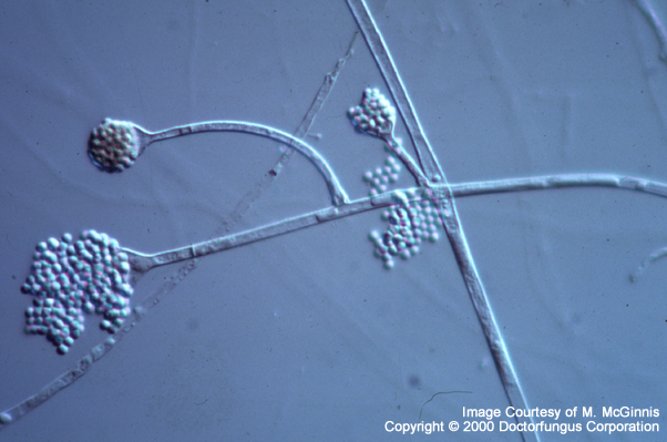
Microscopic Features
Similar to that of the other members of the class Zygomycetes, Absidia corymbifera has wide (6-15 µm in diameter) nonseptate hyphae. A few septa may occasionally be present. Rhizoids are rarely observed. When present, the sporangiophores arise on stolons from points between the rhizoids, but not opposite the rhizoids. The sporangiophores are branched and arise in groups of 2-5 at the internodes. They often produce arches. Sporangiophores carry pyriform, relatively small (20-120 µm in diameter) sporangia. A septum is usually present just below the sporangium in the sporangiophore. The sporangiophore widens to produce the funnel-shaped apophysis beneath the sporangium. The apophysis of Absidia corymbifera is very well-developed and typical. The columella, the tip of the sporangiophore that extends into the sporangium, is semicircular in shape and has a small projection on top. Upon dissolving of the sporangial wall, a short remaining collarette may be observed overlining the apophysis. The sporangiospores are one-celled, hyaline to light black, round to oval in shape, smooth or rarely echinulate on surface and 3-4.5 µm in diameter. They are found in the sporangium and are released to the surrounding when the sporangium ruptures [462, 1295, 2144].
Histopathologic Features
Compare to
The other members of Zygomycetes; Apophysomyces, Mucor, Rhizomucor, and Rhizopus.
Key Features for Differentiation from Other Members of Zygomycetes
| Structure | Differentiation |
|---|---|
| Pyriform sporangium | Absidia (+); Mucor (-), Rhizomucor (-), Rhizopus (-) |
| Septum below the sporangium | Absidia (+); Apophysomyces* (-) |
| Well-developed apophysis | Absidia (+), Mucor (-), Rhizomucor (-), Rhizopus (-) |
| Sporangiophores arising between rhizoids and not opposite them | Absidia (+); Rhizopus (-) |
| Abundant sporulation on routine mycological media | Absidia (+); Apophysomyces (-) |
*has a dark pigmented band below the sporangium
Recent nuclear ribosomal DNA sequence data suggest that Absidia corymbifera is a sister group of Rhizomucor miehei–Rhizomucor pusillus. The studies on molecular phylogeny are still in progress [2341].
Laboratory Precautions
No special precautions other than general laboratory precautions are required.
Susceptibility
In vitro susceptibility data reported so far are limited [1040, 1130]. MIC breakpoints for interpretation of in vitro susceptibility results have not been defined. Similar to the other members of the class Zygomycetes, amphotericin b appears as the sole antifungal drug which is consistently active against Absidia corymbifera . In general, it is resistant to azoles, including the newer derivatives such as voriconazole [2432]. flucytosine is also ineffective against Absidia corymbifera . Some strains may yield relatively low MICs of sordarin group of compounds. However, the significance of this finding is unclear [2212].
In vivo response, on the other hand, largely depends on administration of full-dose amphotericin B therapy as well as extensive surgical debridement and correction of the underlying predisposing factors (such as immunosuppression and diabetic acidosis) [1918].
For MICs of various antifungal drugs so far reported for Absidia, see our N/A(L):susceptibility database.