(described by Carmichael in 1966)
Taxonomic classification
Kingdom: Fungi
Phylum: Ascomycota
Order: Chaetothyriales
Genus: Exophiala
Exophiala jeanselmei
Exophiala spinifera
Description and Natural Habitats
Exophiala is a dematiaceous fungus widely distributed in soil, plants, water, and decaying wood material. As well as being a saprophyte in nature, it is the causative agent of various human infections [531, 1295, 2202].
Species
The genus Exophiala contains several species. The most common ones are Exophiala castellanii, Exophiala jeanselmei, Exophiala moniliae, Exophila pisciphila, Exophiala salmonis, and Exophiala spinifera. Exophiala jeanselmei currently has two varieties: Exophiala jeanselmei var. heteromorpha and Exophiala jeanselmei var. lecanii-corni.
Several morphological and physiological differences aid in differentiation and identification of Exophiala spp. The morphological keys are summarized below.
Key to the Human Pathogenic Species of Exophiala
1. Yeast cells two-celled, usually the predominant form in the culture, rounded at one end, tapering towards the other with annellations at the tapering end; some annellides integrated within hyphae; annelloconidia one-celled, occasionally two-celled, accumulating in balls that tend to slide down the hyphae (Priorly known as Exophiala werneckii and Phaeoannelomyces werneckii and currently reclassified as Hortae werneckii)
1′ two-celled yeast cells absent. (2)
2. Annellides arising on spinelike conidiophores from the hyphae.
2′ Spinelike conidiophores absent. (3)
3. Annellides moniliform with long necks.
3′ Annellides cylindrical to lageniform.
Synonyms
See the summary of synonyms for the Exophiala spp.
Capronia is the probable teleomorphic state of the genus Exophiala [529, 531].
Pathogenicity and Clinical Significance
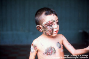
Exophiala spp. are among the fungi causing infections wholly referred to as phaeohyphomycosis. Subcutaneous infections such as mycetoma and chromoblastomycosis may develop due to Exophiala isolates. These infections are usually acquired via traumatic implantation and are associated with the existence of local or systemic immunosuppression, such as organ transplantation. As well as infection and abscess formation in subcutaneous tissues, prosthetic valvular vegetations, fungemia, and disseminated infections due to Exophiala spp. have also been reported. Exophila pisciphila is a neurotropic species causing infections in fish as well as humans [449, 531, 533, 602, 908, 919, 1659, 2184, 2202].
Macroscopic Features
Exophila spp. are initially yeast-like, moist, and brownish to greenish black in color. The texture of the colony eventually becomes velvety due to development of short, aerial grayish hyphae. The front color is olivaceous-black and the reverse is black in mature colonies [531, 1295, 2202].
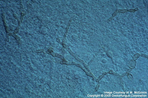
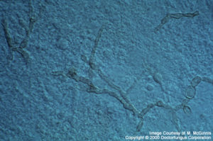
Microscopic Features
In the young cultures, subspherical, budding, yeast-like cells are visualized. These cells often form long chains. As the culture gets older, septate hyphae which bear conidiogenous cells (annelides) are eventually formed. The annelides are tubular and rocket-shaped and typically taper to form a narrow elongated tip. Ellipsoidal, conidia (1-3×3-6 µm) are produced from the annelides. These conidia are usually one-celled and are found in clusters at the apices of annelides or at the sides of the conidiophores [531, 602, 1295, 2202].
Histopathologic Features
Phaeoid (brown) hyphae and phaeoid yeast-like cells are visualized in infected tissues [462].
Compare to
Phialophora
Phaeoannellomyces
Wangiella
Exophiala species differ from members of the genera Phialophora and Wangiella by forming annellides rather than phialides. Variations in decomposition of casein and tyrosine, ability to grow in 15% sodium chloride, KNO3 assimilation, and the maximum temperature of growth also help in differentiation of Exophiala from Phaeoannellomyces and Wangiella [1295].
Some of the species have synanamorphic forms in the genera Cladosporium, Phialophora, Rhinocladiella, Phaeococcomyces and Phaeoannellomyces.
Laboratory Precautions
No special precautions other than general laboratory precautions are required.
Susceptibility
There are a small number of reports available on a limited number of isolates. The clinical significance of these data remains unclear [384, 531, 558, 1490, 1491, 1864].
For Exophiala jeanselmei, fluconazole yielded very high MICs. The MICs of flucytosine and miconazole appeared lower than fluconazole but were still relatively high. MICs of amphotericin B, ketoconazole, and voriconazole were similar and relatively low. The lowest MICs were obtained for itraconazole and terbinafine. Exophiala jeanselmei showed decreased susceptibility to both amphotericin B deoxycholate and ABLC in other workers’ hands. The novel echinocandin, caspofungin, on the other hand, yielded favorable in vitro activity against Exophiala jeanselmei isolates.
For Exophiala pisciphila, amphotericin B yielded the highest MICs. Flucytosine and voriconazole MICs were similar; itraconazole and ketoconazole MICs were lowest.
For Exophiala spiniphera, MICs of amphotericin B, itraconazole, and voriconazole were similar and encouragingly low.
For MICs of various antifungal drugs for Exophiala, see our N/A(L):susceptibility database.
The correlation of in vitro susceptibility with clinical outcome for Exophiala is not yet known. While there is no standard treatment, combination of surgical and antifungal treatment is usually preferred for subcutaneous Exophiala infections. Amphotericin B with or without flucytosine, itraconazole, and terbinafine have been used. The combination of amphotericin B, flucytosine and itraconazole appeared useful in a case with subcutaneous infection due to Exophiala jeanselmei [449, 462, 919, 2184, 2425].