(described by Lechevalier and Lechevalier in 1970)
Taxonomic Classification
Actinomadura is an aerobic actinomycetes.
Description and Natural Habitats
Actinomadura is a filamentous bacterium found in soil. Although it was once believed to be a fungus, the information later attained about its ultrastructural cellular properties showed that Actinomadura is in fact an aerobic actinomycetes. However, for the reason that most of the diagnostic procedures related to Actinomadura are still held in mycology laboratories in many centers, the discussion on Actinomadura is included in this website.
Species
Actinomadura madurae, Actinomadura pelletieri, and Actinomadura dassonvillei are the species included in the genus Actinomadura. Actinomadura madurae is distinguished from A. pelletieri by its ability to produce acid from cellobiose.
Pathogenicity and Clinical Significance
It is one of the common causes of actinomycotic mycetoma (maduramycosis or madura foot), characterized by formation of granules containing branched filaments. It is a chronic infection involving the subcutaneous tissue, muscles and bones particularly of lower extremities. The characteristic granules formed during the course of infection drain via formation of sinuses.
Macroscopic Features
The growth rate of Actinomadura is slow. It grows on routine mycologic or mycobacteriologic media and under aerobic conditions. The colony has a glabrous, waxy, membranous or mucoid, heaped and folded appearance. The color of the colony is red, pink, yellow, orange, white, or tan. Following two weeks of incubation, aerial hyphae may develop on the surface, particularly on Lowenstein-Jensen medium [1295].
Microscopic Features
Actinomadura is Gram positive and nonacid-fast. The typical structures are the filaments which are nonfragmenting and narrow (0.5-1 µm in diameter). These filaments branch abundantly. Chains of round conidia may occasionally be produced from the aerial hyphae, particularly on slide cultures [1295].
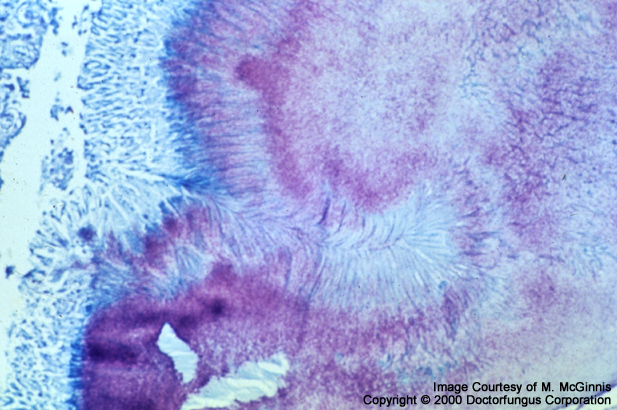
Histopathologic Features
The granules of Actinomadura are 0.1 to several mm in diameter. They are composed of branching, beaded, Gram positive filaments which are delicate and around 1 µm in diameter [462].
Compare to
Nocardia, Nocardiopsis, and Streptomyces.
Key Features for Differentiation
Some of the properties related to the microscopic features and growth of the aerobic actinomycetes are summarized in the Table below. The color and texture of the colony, hydrolysis of casein, tyrosine, xanthine, gelatin, starch, and urea, and acid formation from lactose, xylose, and cellobiose should also be examined for definitive identification of these bacteria at species level [1295].
| STRUCTURE/PROPERTY | DIFFERENTIATION |
|---|---|
| Acid fast staining | Actinomadura (-) Nocardiopsis (-) Streptomyces* (-) Nocardia (+) |
| Spontaneous fragmentation of hyphae | Actinomadura (-) Streptomyces (-) Nocardia (+) Nocardiopsis (+) |
| Ability to grow in existence of lysozyme | Actinomadura (-) Nocardiopsis (-) Streptomyces** (-) Nocardia (+) |
*The spores of some Streptomyces spp. may be acid fast.
**Some strains may grow.
Laboratory Precautions
No special precautions other than general laboratory precautions are required.
Susceptibility
Treatment regimens usually consist of a combination therapy. Since mycetoma may be caused by several different fungi or aerobic actinomycetes, identification of the causative agent and differentiation of eumycotic mycetoma from actinomycotic mycetoma constitute the major points in application of appropriate antimicrobial therapy. While Actinomadura madurae is usually suceptible to a combination of streptomycin and dapsone, Actinomadura pelletieri responds better to a combination of streptomycin and trimethoprim-sulfamethoxazole. As well as antimicrobial therapy, surgery (limited and bulk-reduction surgery, preferentially) is required for some patients.