Berkhout, 1923 nom. cons.
(described by Bennett in 1844)
Taxonomic classification
Kingdom: Fungi
Phylum: Ascomycota
Subphylum: Ascomycotina
Class: Ascomycetes
Order: Saccharomycetales
Family: Saccharomycetaceae
Genus: Candida
Description and Natural Habitats
Candida is a yeast and the most common cause of opportunistic mycoses worldwide. It is also a frequent colonizer of human skin and mucous membranes. Candida is a member of normal flora of skin, mouth, vagina, and stool. As well as being a pathogen and a colonizer, it is found in the environment, particularly on leaves, flowers, water, and soil. While most of the Candida spp. are mitosporic, some have known teleomorphic state and produce sexual spores. See the page on individual species for more detailed information on teleomorphic genera.
Species
The genus Candida includes around 154 species. Among these, six are most frequently isolated in human infections. While Candida albicans is the most abundant and significant species, Candida tropicalis, Candida glabrata, Candida parapsilosis, Candida krusei, and Candida lusitaniae are also isolated as causative agents of Candida infections. Importantly, there has been a recent increase in infections due to non-albicans Candida spp., such as Candida glabrata and Candida krusei [4, 29, 117]. Patients receiving fluconazole prophylaxis are particularly at risk of developing infections due to fluconazole-resistant Candida krusei and Candida glabrata strains [186]. Nevertheless, the diversity of Candida spp. that are encountered in infections is expanding and the emergence of other species that were rarely in play in the past is now likely [158, 259, 288, 294].
| Species Commonly Causing Invasive Candidiasis | |
| Species | Frequency |
|---|---|
| Candida albicans Candida tropicalis Candida glabrata Candida parapsilosis Candida krusei Candida lusitaniae |
50% 15-30% 15-30% 15-30% ~1% ~1% |
Synonyms
See the summary of synonyms and teleomorph-anamorph relations for the Candida spp.
Infections caused by Candida spp. are in general referred to as candidiasis. The clinical spectrum of candidiasis is extremely diverse. Almost any organ or system in the body can be affected. Candidiasis may be superficial and local or deep-seated and disseminated. Disseminated infections arise from hematogenous spread from the primarily infected locus. Candida albicans is the most pathogenic and most commonly encountered species among all. Its ability to adhere to host tissues, produce secretory aspartyl proteases and phospholipase enzymes, and transform from yeast to hyphal phase are the major determinants of its pathogenicity. Several host factors predispose to candidiasis [242, 266, 270]:
| PREDISPOSING FACTOR | EXAMPLES |
|---|---|
| Physiological | Pregnancy, age (elderly and infancy) |
| Trauma | Maceration, infection, burn wound |
| Hematological | Neutropenia, cellular immunodeficiency (leukemia, lymphoma, AIDS, aplastic anemia) |
| Endocrinological | Diabetes Mellitus, hypoparathyroidism, Addison’s disease |
| Iatrogenic | Chemotherapeutics, corticosteroids, oral contraceptives, antibiotics, catheters, surgery |
| Other | Intravenous drug addiction, malnutrition, malabsorption, thymoma |
Candidiasis is mostly an endogenous infection, arising from overgrowth of the fungus inhabiting in the normal flora. However, it may occasionally be acquired from exogenous sources (such as catheters or prosthetic devices) [167] or by person-to-person transmission (such as oral candidiasis in neonates of mothers with vaginal candidiasis or endophthalmitis following corneal transplantation from an infected donor) [210].
Macroscopic and Microscopic Features
The colonies of Candida spp. are cream colored to yellowish, grow rapidly and mature in 3 days. The texture of the colony may be pasty, smooth, glistening or dry, wrinkled and dull, depending on the species [1295].
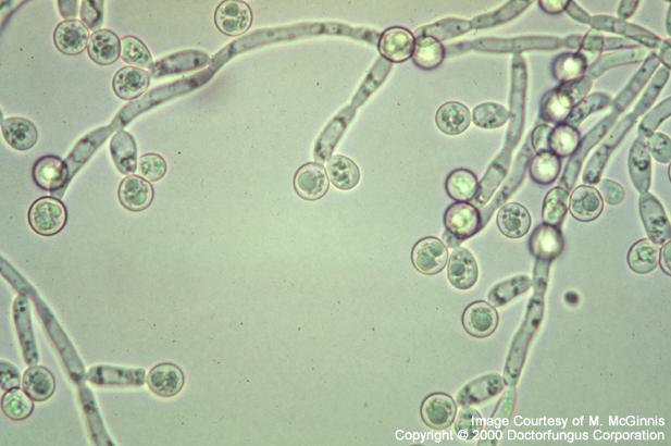
The microscopic features of Candida spp. also show species-related variations. All species produce blastoconidia singly or in small clusters. Blastoconidia may be round or elongate. Most species produce pseudohyphae which may be long, branched or curved. True hyphae and chlamydospores are produced by strains of some Candida spp.
Although they are the members of the same genus, the various species do have some degree of unique behavior with respect to their colony texture, microscopic morphology on cornmeal tween 80 agar at 25°C (Dalmau method) and fermentation or assimilation profiles in biochemical tests [1295].
Choose a species for more detail:
- Candida albicans
- Candida tropicalis
- Candida glabrata
- Candida parapsilosis
- Candida krusei
- Candida lusitaniae
Histopathologic Features
See our histopathology page.
Compare to
Candida spp. should be differentiated from other clinically encountered yeasts, such as Blastoschizomyces, Cryptococcus, Geotrichum, Malassezia, Rhodotorula, Saccharomyces, and Trichosporon. Morphology on cornmeal tween 80 agar, capsule production, urease activity, ability to grow in presence of cycloheximide, growth pattern in Sabouraud broth, and fermentation assimilation profiles help in differentiation of Candida from other yeasts. Well-developed pseudohyphae and one-celled blastoconidia characterize the common species of Candida. Candida differs from Cryptococcus by having well-developed pseudohyphae. The lack of arthroconidia is the major microscopic feature which differentiates Candida from Trichosporon and Geotrichum, the two genera that produce abundant arthroconidia [1295].
Laboratory Precautions
No special precautions other than general laboratory precautions are required.
Susceptibility
Please see our extended discussion of the usual susceptibility patterns of Candida spp.