Synonyms
African histoplasmosis. Contrast with North American histoplasmosis due to Histoplasma capsulatum
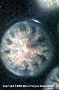
Yeast Phase of Histoplasma capsulatum var duboisii, GMS stain
Definition and forms of the disease
Histoplasmosis due to N/A(L):Histoplasma duboisii is a mycotic infection primarily involving cutaneous, liver, lung, lymphatic, subcutaneous, and bony tissues. Skin and bone are the most frequently invaded sites [454]. The etiologic agent grows as a large yeast within giant cells. It may also present with small cells that are typical of those seen in histoplasmosis due to Histoplasma capsulatum. Nodular and ulcerative cutaneous and osteolytic lesions of bone that disseminate or remain localized are the primary clinical characteristics of histoplasmosis duboisii [11, 1185, 1186, 2101].
Prognosis and therapy
Special resource: You may also want to refer to the Infectious Disease Society of America-Mycoses Study Group (IDSA-MSG) Practice Guidelines for this disease. It is available at the (E):IDSA website.
Isolated lesions may heal spontaneously. Surgical management is also an option. Disseminated disease has a grave prognosis, especially if the liver and spleen are involved. Amphotericin B, itraconazole or fluconazole are the drugs of choice [6, 36, 1690, 2302].
Histopathology
Minimal cellular reaction to the fungi is noted with the exception of large numbers of giant cells (up to 80 µm) and macrophages. Neutrophils are usually present, especially during necrosis. The globose to ovoid, thick-walled yeasts are 7-15 µm (average 10 µm) in diameter and may form rudimentary pseudohyphae consisting of 4 or 5 cells. Large aggregates of yeast cells can be readily seen within giant cells and extracellularly following necrosis of the host tissue. Unlike N/A(L):Blastomyces dermatitidis, the blastoconidia are not attached to the parent cell by a broad neck.
Laboratory
Direct examination
Clinical specimens such as tissue are examined in 10% KOH or calcofluor. The large yeast cells should be readily visible. Care must be taken to ensure that B. dermatitidis is not confused with the etiologic agent of histoplasmosis duboisii since they both occur in Africa.
Isolation
Inoculate the clinical material onto Inhibitory Mould Agar and/or yeast extract-phosphate agar and/or BHI agar with 10% sheep blood and/or a medium containing cycloheximide. Incubate cultures at 30°C and do not discard until 12 weeks.
Laboratory confirmation
Confirmation is necessary to ensure that the fungus is not a species of N/A(L):Chrysosporium or N/A(L):Sepedonium. This can be accomplished by the mould to yeast conversion, exoantigen technique or DNA probes. The etiologic agents of histoplasmosis capsulati and histoplasmosis duboisii are morphologically identical at 30°C and by DNA testing and exoantigen confirmation.