Ajello, 1959
Macroscopic morphology
The growth rate of Microsporum cookei is moderately rapid. The diameter of the colony reaches 1 to 3 cm following incubation at 25°C for 7 days on Sabouraud dextrose agar. The texture is downy to powdery. The front color is initially white to yellow and may turn to grape red or dark brown in time. The reverse is dark red to dark brown.
Microscopic morphology
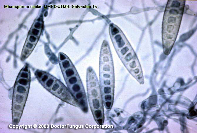
Microsporum cookei produces septate branching hyphae, macroconidia, and microconidia. The macroconidia are numerous, oval in shape, thick-walled, rough and 6- to 10-celled. Microconidia are also abundant. They are unicellular and ovoid to pyriform in shape.
Special notes
In vitro hair perforation test is positive. The thick-walled macroconidia and the dark red pigment of Microsporum cookei differentiate it from Microsporum gypseum. The teleomorph of Microsporum cookei is called Arthroderma cajetani.
While Microsporum cookei is known to produce infection in rodents, its association with tinea corporis in humans is rare and perhaps doubtful.