Fuentes, 1956
Macroscopic morphology
Microsporum nanum grows moderately rapidly and the diameter of the colony reaches 1 to 3 cm following incubation at 25°C for 7 days on Sabouraud dextrose agar. The texture is powdery, cottony, thin, spreading, velvety or flat and often has some radial, shallow furrows. The color is white to dark beige from the front and reddish brown from the reverse.
Microscopic morphology
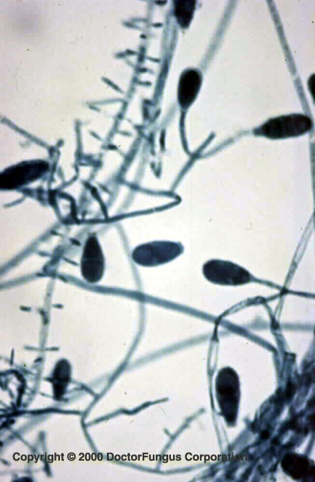
Microsporum nanum produces septate hyphae, macroconidia, and microconidia. Macroconidia are 1- to 4-celled (usually 2) and thin walled and oval to elliptical in shape. Microconidia are club-shaped and their abundance may vary.
Special notes
In vitro hair perforation test is positive. No special growth factor is required to grow Microsporum nanum. Its teleomorph is called Arthroderma obtusa. Microsporum nanum is a rare cause of tinea corporis in humans and a frequent cause of ringworm in its natural reservoir, the pig.
Microsporum nanum requires differentiation from Trichothecium spp. and Chrysosporium spp. Unlike Chrysosporium spp., Microsporum nanum produces microconidia. Unlike Trichothecium spp., it produces macroconidia that are solitary, but not in clusters, and it is resistant to cycloheximide.