Georg, Ajello, Friedman et Brinkman, 1962

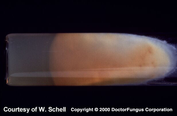
Sabouraud dextrose agar (SDA), reverse of the colony
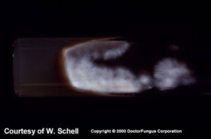
SDA, front of the colony
Macroscopic morphology
Microsporum vanbreuseghemii colonies grow moderately rapidly and mature in a week. The texture is flat, spreading and granular to velvety. The front color is cream yellow, lavender pink or tan. A lemon yellow or yellow to orange diffusible pigment is observed from the reverse.
Microscopic morphology
Microsporum vanbreuseghemii produces septate branching hyphae, macroconidia, and microconidia. Macroconidia are 6- to 13-celled, cylindrical (pencil-shaped) to fusiform, rough, and thick-walled. Microconidia are unicellular and obovoid to pyriform in shape.
Special notes
In vitro hair perforation test is positive. The teleomorph state of Microsporum vanbreuseghemii is called Arthroderma grubyi. It mostly infects cats, dogs, and squirrels. It is a rare cause of tinea corporis in man.
[1295]