(described by Bainier in 1907)
Taxonomic classification
Kingdom: Fungi
Phylum: Ascomycota
Class: Euascomycetes
Order: Eurotiales
Family: Trichocomaceae
Genus: Paecilomyces
Description and Natural Habitats
Paecilomyces is a cosmopolitan filamentous fungus which inhabits the soil, decaying plants, and food products. Some species of Paecilomyces are isolated from insects. The telemorphs of Paecilomyces are classified in the genera Byssochlamys, Chromocleista, Talaromyces, and Thermoascus [531]. Paecilomyces is usually considered as a contaminant but may also cause infections in humans and animals.
Species
The genus Paecilomyces contains several species. The most common are Paecilomyces lilacinus and Paecilomyces variotii. The color of the colony and certain microscopic features help in differentiation of the Paecilomyces species from each other [531]. Another feature that helps in species identification is thermophilicity. Paecilomyces crustaceus and Paecilomyces variotii are thermophilic and can grow well at temperatures as high as 50° and possibly 60°C [531, 2202].
See the list of obsolete names, synonyms, and telemorphs for Paecilomyces spp.
Pathogenicity and Clinical Significance
Paecilomyces species can cause various infections in humans. These infections are occasionally referred to as paecilomycosis. Corneal ulcer, keratitis, and endophthalmitis [1772] due to Paecilomyces may develop following extended wear contact lens use or ocular surgery. Paecilomyces is among the emerging causative agents of opportunistic mycoses in immunocompromised hosts [908]. Direct cutaneous inoculation may lead to these infections [1695]. These infections may involve almost any organ or system of human body. Soft tissue [2437], pulmonary [355], and cutaneous infections [1695, 1998], cellulitis [1110], onychomycosis [746], sinusitis [923, 1938], otitis media [585], endocarditis [969, 1476, 2109, 2274], osteomyelitis [456], peritonitis [482, 1238, 1928], and catheter-related fungemia [412, 2221] have all been reported. Paecilomyces species can also cause allergic disorders, such as allergic alveolitis [33].
Paecilomyces can cause infections in animals as well. It has been reported as a cause of hyalohyphomycosis in various animals, such as cats [663], laboratory rats [1265], turtles [1833], and goats [1754].
Macroscopic Features
Colonies of Paecilomyces grow rapidly and mature within 3 days. Paecilomyces crustaceus and Paecilomyces variotii are thermophilic and can grow well at temperatures as high as 50° and possibly 60°C. The colonies are flat, powdery or velvety in texture. The color is initially white, and becomes yellow, yellow-green, yellow-brown, olive-brown, pink, or violet, depending on the species. The reverse is dirty white, buff or brown. A sweet aromatic odor may be associated with older cultures [531, 1295, 2144].
Microscopic Features
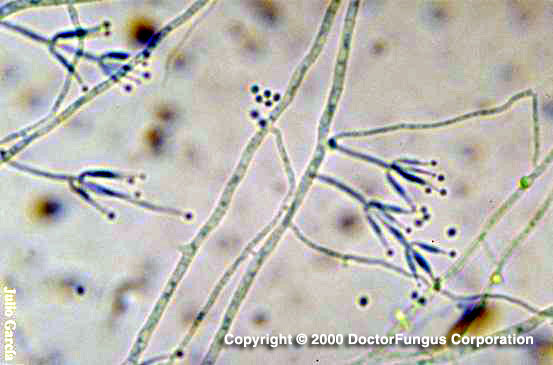
Septate hyaline hyphae, conidiophores, phialides, conidia, and chlamydospores are observed. Conidiophores (3-4 µm wide and 400-600 µm long) are often branched and carry the phialides at their tips. The phialides are swollen at their bases and taper towards their apices. They are usually grouped in pairs or brush-like clusters. Conidia are unicellular, hyaline to darkly colored, smooth or rough, oval to fusoid, and form long chains. Chlamydospores are occasionally present [1295, 2202].
Ascomata, asci, and ascospores are produced by the telemorphic species, such as Thermoascus crustaceus [531].
Histopathologic Features
Septate hyphae may be observed. See also our histopathology page.
Compare to
Phialides of Paecilomyces are basally swollen, taper towards their apices and are organized slighly apart from eachother. Phialides of Penicillium, on the other hand, have thicker apices and are organized in tight clusters. Colonies of Penicillium are commonly blue-green in color while those of Paecilomyces are not.
Laboratory Precautions
No special precautions other than general laboratory precautions are required.
Susceptibility
Very limited data are available and these data include Paecilomyces fumosoroseus, Paecilomyces javanicus, Paecilomyces lilanicus, Paecilomyces marquandii, and Paecilomyces variotii. The MICs of amphotericin B tend to be low, except for strains of Paecilomyces lilanicus. On the other hand, the MICs of flucytosine tend to be very high, except for strains of Paecilomyces variotii. Fluconazole yields considerably high MICs for all species tested. Itraconazole and ketoconazole MICs are lowest for Paecilomyces variotii compared to other species [23]. Voriconazole has been tested against Paecilomyces lilanicus and Paecilomyces variotii and has promisingly low MICs for both of these species [687, 2432]. Posaconazole [1434], the novel triazole UR-9825 [374], and terbinafine
[2308] appear active in vitro against Paecilomyces. Caspofungin, on the other hand, is active in vitro against Paecilomyces variotii but lacks meaningful activity against Paecilomyces lilanicus [558, 1780].
For MICs of various antifungal drugs for Paecilomyces, see our N/A(L):susceptibility database.
Despite high MICs in vitro, amphotericin B and flucytosine combination has been succesfully used in treatment of some cases with paecilomycosis due to Paecilomyces lilanicus [412, 1110]. Caspofungin combined with itraconazole has also been found to be successful in a case infected with Paecilomyces lilanicus [1998].