Tinea corporis, (A):Tinea cruris, and Tinea pedis
Tinea corporis
Synonyms
Ringworm of the body
Tinea corporis
Ringworm
Tinea manuum
Definition
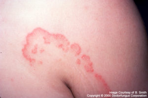
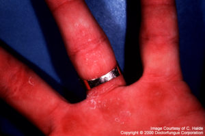
The key characteristic of Tinea corporis is that the fungus involves the glabrous (relatively hairless) skin. The infection is limited to the stratum corneum of the epidermis. Vellus hair (the fine hair present on glabrous skin) may be invaded, and the hair follicle may serve as a reservoir for the fungus. Tinea pedis,Tinea manuum, and (A):Tinea cruris refer to Tinea corporis that is limited to the foot, hand, and groin, respectively. There is otherwise little special about them.
History lesson: The term tinea has an interesting origin. A worm of a moth would sometimes grow on a woolen blanket. The resulting round holes were similar to the rounded lesions seen on the skin of patients. The genus name for the moth was Tinea, and thus this name was used as part of the Latin binomials naming these infections.
Epidemiology
Transmission of tinea corporis may occur from direct contact with infected animals (especially cats and dogs), infected humans, or contaminated fomites such as furniture and clothing. Like many other fungal skin infections, warmth and humidity favor the occurrence of this infection. Therefore, tropical and subtropical regions have a higher incidence of tinea corporis [1447].
Tinea imbricata is an unusual form of Tinea corporis caused by T. concentricum. This form of Tinea is characterized by ring-like growth in overlapping circles that may have an autosomal dominant genetic predisposition [1878, 2080]. This may explain its geographic restriction to certain regions of the Far East, South Pacific, and South and Central America [335, 1009].
Clinical manifestations
Tinea corporis can present on any area of the body. Zoophilic organisms commonly affect exposed areas like the face, neck and arms. Oppositely, anthropophilic organisms classically affect occluded areas of the skin or areas of trauma. In regards to the clinical appearance, multiple varieties have been described, and to make things a little more confusing, many of them have distinct names although they are all forms of tinea corporis![1447].
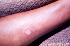
The classical and more common clinical variety in which annular lesions have active, erythematous and spreading borders with central clearing is called in common parlanceringworm and scientifically,tinea circinata. When herpetiform, subcorneal vesicles appear the term “bullous tinea corporis” has been used.
Between 3 to 4% of tinea corporis cases present with an erythematous, scaly rash on the face with or without telangiectasia, atrophy, and photoexacerbation. This clinical form, calledtinea fasciale, may be confused with lupus erythematosis [838, 1931].
When any of these dermatophytoses are treated with corticosteroids, the lesions take on an atypical appearance and lose the characteristic scaling of tinea corporis. Patches, papules, or small nodules appear. For this variety the termtinea incognito is used [1103].
Tinea profunda refers to the appearance of subcutaneous abscesses, which are frequently associated with T. mentagrophytes.
Tinea axillaris refers to the involvement of the axillary (armpit) region [213].Other atypical inflammatory forms include the appearance of verrucous lesions, kerion-like lesions and/or nodular granulomas (Majocchi’s granuloma).
Prognosis and therapy
N/A(L):Topical therapy is useful in cases of noninflammatory lesions. Systemic therapy with griseofulvin, terbinafine, ketoconazole, itraconazole or fluconazole is recommended for inflammatory varieties [616]. The following regimens are recommended:
| Drug | Regimen | Reference |
|---|---|---|
| Griseofulvin | 500 mg once daily for 4-6 weeks | [703] |
| Fluconazole | 150 mg once daily for 4-6 weeks | [703] |
| Itraconazole | 100 mg daily for 1-2 weeks | [1721] |
| Terbinafine | 250 mg daily for 2-4 weeks | [715] |
Histopathology
Rarely is a biopsy required for the diagnosis of this condition. Classically, fungal elements are seen in the stratum corneum with PAS, silver methenamine or other fungal stain. If they are absent, pathology will show nonspecific lesions that resemble acute or chronic dermatitis [1447].
Laboratory
Direct examination
Superficial scraping from the spreading border of the lesion is recommended. If vesicular lesions predominate, the roof of the blister should be taken. These samples are mounted in 10% KOH and septate, branching hyphae with or without arthrospores should be seen.
Inoculate the clinical specimens onto Sabouraud glucose agar, incubate at 30°C and discard negative cultures in 4 weeks. For more details consult the chapters on the three species involved: Epidermophyton, Trichophyton, and Microsporum.
Mycology (principal dermatophytes)
The three most common responsible for tinea corporis are:
- Trichophyton rubrum
- Microsporum canis
- Trichophyton mentagrophytes
However, other species implicated include:
- Epidermophyton floccosum
- Microsporum audouinii
- Microsporum gypseum
- Trichophyton megninii
- Trichophyton schoenleinii
- Trichophyton tonsurans
- Trichophyton verrucosum
Natural habitat
Animals, humans, soil
Tinea cruris
Synonyms
Jock itch, ringworm of the groin
Definition
Tinea cruris is an acute or chronic infection of the groin, perineum, and perianal region.
Epidemiology
This dermatophytoses is more commonly seen in men. According to Martin et al., the apparent reasons for this include [1447]:
- The temperature, humidity, and occlusion of the scrotum and groin area, especially related to the clothing, are ideal for the development of these fungi.
- Men suffer more frequently from other dermatophytoses, particularly (A):tinea pedis, and cross infection between sites is very common.
Both direct contact between infected individuals and indirect contact with nonliving contaminated objects (towels, clothing, bed linens, urinals, and bed pans) are ways of transmission. Tropical climates and summer months in temperate regions appear to promote higher rates of this infection.
Clinical manifestations
Tinea cruris presents with sharply demarcated lesions with a raised erythematous margin and thin dry epidermal scaling. Papulovesicular lesions may also be present but pustules such as those caused by Candida are very unusual. Lesions classically involve the genitocrural area and medial upper thigh in a symmetrical fashion, but asymmetrical involvement may occur. The scrotum is usually minimally affected, and this is a distinct contrast with infections of this area by Candida (“Intertrigo”). Extension to the pubic area, lower abdomen, buttock, and perianal areas occurs rarely but can be seen, especially if Trichophyton rubrum is the causative agent [1447]. Patients complain initially of intense pruritus, but the lesions will become painful if maceration and superinfection occur. In addition to candidiasis (“intertrigo”), the differential diagnosis also includes lichen simplex and erythrasma.
Prognosis and therapy
N/A(L):Topical therapy is usually enough to cure tinea cruris. Drugs formulated in powders or minimally occlusive cream bases are preferred. Oral antifungal agents such as griseofulvin, terbinafine, ketoconazole, itraconazole or fluconazole are reserved for widespread and severely inflamed cases [616]. Recommended regimens are (A):the same discussed for Tinea corporis.
Rates of relapse for this infection are very high, therefore hygiene measures are crucial for long term success. They should include thorough drying, the use of well-ventilated clothing, and separate towels for the groin area.
Histopathology and laboratory
As mentioned above, tinea cruris is just an anatomic variety of tinea corporis, therefore pathologic and diagnostic considerations are the same.
Mycology (principal dermatophytes)
- Epidermophyton floccosum
- Microsporum canis
- Trichophyton mentagrophytes
- Trichophyton rubrum
Natural habitat
Humans
Tinea pedis
Synonyms
Athlete’s foot, ringworm of the foot
Tinea pedis
Definition
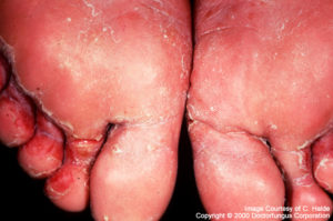
Tinea pedis is a fungal infection of the feet, principally involving the toe webs and soles.
Epidemiology
This infection is related to footware and is considered to been a “new” dermatophytoses in that it began in association with use of footware [1791]. More occlusive shoes are associated with higher chances of having tinea pedis. Contact with bath or pool floors is another recognized risk factor and the rate of infection increases in relation with the number of people using the facilities. The infection is more common during summer months and in tropical climates [1447].
Clinical manifestations
Tinea pedis may present in several ways, varying from mild chronic scaling to acute lesions that are exfoliative, pustular, or bullous. Four general clinical forms can be distinguished [1447]:
- Intertriginous tinea pedis: this form typically affects the toe webs and subdigital areas producing chronic scaling, fissuring and maceration. The 4th to 5th and 3rd to 4th interdigital areas are the most commonly affected. Some studies have implicated the interaction of dermatophytes with the mixed skin flora characteristic of this area in the pathogenesis of this infection [1336].
- Tinea pedis with a papulosquamous pattern (“moccasin-like”): as the name suggests, this form affects the soles and lateral aspects of the feet in a pattern suggestive of the skin covered were one wearing a moccasin. Scaling is the main process while inflammation is minimal. This type is usually bilateral and interestingly may present with concomitant involvement of one hand in a pattern called “one hand, two feet”.
- Vesicular or vesiculobullous tinea pedis: this form involves the instep and the mid-anterior aspect of the sole. It causes small, confluent vesicles or vesiculopustules surrounded by scaling. Scales may extend to the toewebs. Occasionally, large bullae appear.
- Ulcerative tinea pedis: this type causes a more acute and symptomatic picture characterized by maceration and ulceration of large areas of the soles. White hyperkeratosis and a strong heady odor are characteristic. Bacterial superinfection, usually with gram-negative organisms, is frequent and should be taken into account at the time of treating this condition.
Despite the previously detailed characterizations, it is not unusual to see a presentation in which there is overlap of two and even three of these clinical varieties. A correlation between lower extremity erysipelas and tinea pedis has also been established [1953].
Prognosis and therapy
Even though N/A(L):topical antifungals can be used to treat mild, noninflammatory varieties of tinea pedis, systemic therapy is helpful to treat hyperkeratotic areas such as the soles [616]. Terbinafine and itraconazole have proven to be effective [1320]. Several effective regimens have been described:
| Drug | Regimen | Reference |
|---|---|---|
| Terbinafine | 250 mg daily for 2 weeks | [534, 1008] |
| Itraconazole | 100 mg daily for 4 weeks 200 mg daily for 2 weeks 200 mg BID for 1 week |
[534, 944, 1008] |
The experience with fluconazole is minimal. Interestingly with respect to the not infrequent bacterial superinfection seen with this form of dermatophytoses, antibacterial properties have been described for terbinafine [1650] and might contribute to its success as a therapy.
Histopathology
Rarely is a biopsy required for the diagnosis of this condition. Classically, fungal elements are seen in the stratum corneum with PAS, silver methenamine or other fungal stain. If they are absent, pathology will show nonspecific lesions of acute or chronic dermatitis [1447].
Laboratory
Direct examination
Superficial scraping from the spreading border of the lesion is recommended. If vesicular lesions predominate, the roof of the blister should be taken. These samples are mounted in 10% KOH and septate, branching hyphae with or without arthrospores should be seen.
Isolation
Inoculate the clinical specimens onto Sabouraud glucose agar, incubate at 30°C and discard negative cultures in 4 weeks. For more details consult the chapters on the three species involved: N/A(L):Epidermophyton, Trichophyton, and Microsporum.
Mycology (principal dermatophytes)
- N/A(L):Epidermophyton floccosum
- Trichophyton mentagrophytes
- Trichophyton rubrum
- Trichophyton tonsurans
Natural habitat
Humans, animals