Behrend (1890)
Macroscopic morphology
Colonies on Sabouraud dextrose agar at 25°C are white, dry, and farinose with irregular folds. Colony size is 10-13 mm after 7 days incubation.
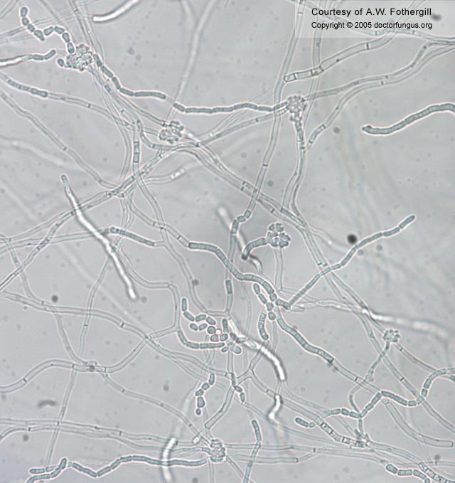
Microscopic morphology
On cornmeal following 72 hours incubation at 25°C, it produces true hyphae that disarticulate into rectangular arthroconidia measuring approximately 3-5 x 4-16 µm. Appressoria are formed[928, 1270].
Special notes
This isolate is urease positive with growth on media containing cycloheximide, has variable growth at 37°C, and fails to grow at 42°C. The type strain was isolated from the hair of a patient with white piedra of the scalp. This species is most frequently associated with white piedra of the scalp but has also been recovered from wounds and CSF. Trichosporon ovoides may be distinguished from T. asahii by formation of appressoria and from T. inkin by presence of a marginal zone, positive growth on cycloheximide and its failure to develop sarcinae (septations within the arthroconidia). Source of infection is also important in separating the species with T. ovoides being primarily isolated from white piedra of the scalp and T. inkin being primarily implicated in white piedra of the groin[928]. It has also been implicated in summer-type hypsersensitivity penumonitis[2188].