(described by Corda in 1833)
Taxonomic Classification
Kingdom: Fungi
Phylum: Ascomycota
Class: Euascomycetes
Order: Onygenales
Family: Onygenaceae
Genus: Chrysosporium
Chrysosporium spp.
Chrysosporium xerophilum
Emmonsia parva
Description and Natural Habitats
Chrysosporium is a keratinophilic filamentous fungus commonly isolated from soil, plant material, dung, and birds. It lives on remains of hairs and feathers in soil. The telemorphs of Chrysosporium spp. are included in the genera Aphanoascus, Nannizziopsis, and Uncinocarpus. As well as being a common contaminant, Chrysosporium is occasionally isolated from human infections.
Species
The genus Chrysosporium contains several species. The most common ones are Chrysosporium keratinophilum, Chrysosporium tropicum, Chrysosporium merdarium, Chrysosporium inops, Chrysosporium pannicola, Chrysosporium queenslandicum, and Chrysosporium zonatum. Another species of special interest, classified as Emmonsia parva on the ecological basis, is occasionally named as Chrysosporium parvum as well. Please see our detailed discussion about the classification of Emmonsia parva.
The species of Chrysosporium are differentiated from each other by the texture of the colony and morphology, location, and size of the conidia. Also, some species, particularly Chrysosporium pannicola, do not grow at 37°C [531].
See the summary of synonyms and telemorph-anamorph relations for the Chrysosporium spp.
Pathogenicity and Clinical Significance
Chrysosporium spp. may cause skin infections and onychomycosis in humans. In addition to these superficial infections, Chrysosporium spp. have occasionally been isolated from systemic infections in bone marrow transplant recipients and in patients with chronic granulomatous disease [1581, 1952]. The high mortality rate of systemic Chrysosporium infections is noteworthy [1581].
Macroscopic Features
Chrysosporium colonies grow moderately rapidly at 25°C. The morphology of the colonies is very variable. They may be granular, woolly, or cottony and flat, or raised and folded in appearance. From the front, the color is white cream, yellow or tan to pale brown. The reverse is white to brown [531, 1295, 2202].
Microscopic Features
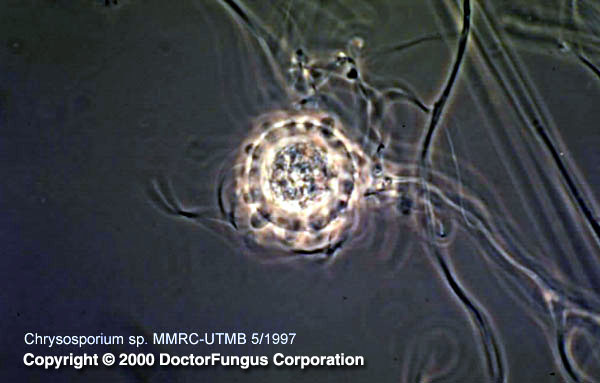
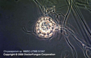
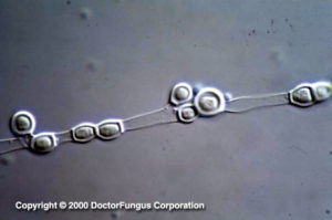
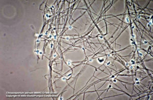
Chrysosporium produces hyphae, conidia (aleuriconidia), and arthroconidia. Hyphae are septate while the conidia are hyaline, broad-based, one-celled, and smooth- or rough-walled. These conidia are broader than the vegetative hyphae and occur terminally on pedicels, along the sides of the hyphae, or in intercalary positions. The conidia usually have an annular frill which is the reminant of the hyphal wall that remains after detachment from the hypha. Arthroconidia, on the other hand, are abundant and larger than their parent hyphae in diameter. In addition, Chrysosporium parvum forms enlarged, thick-walled cells (adiaspores) at 37-40°C [531, 1295, 2202].
Compare to
(A):Blastomyces dermatitidis
Geomyces spp.
Microsporum spp.
Sepedonium spp.
Trichophyton spp.
Chrysosporium differs from Blastomyces by not being dimorphic; from Microsporum and Trichophyton by lacking macroconidia; from Geomyces by lacking branched, fertile hyphae on erect conidiophores; and from Sepedonium by having hyaline conidia.
Laboratory Precautions
No special precautions other than general laboratory precautions are required.
Susceptibility
Very limited data are available. Amphotericin B, itraconazole, ketoconazole, and voriconazole tend to yield low MICs for Chrysosporium keratinophilum isolates. Fluconazole MICs are relatively higher than those of the just noted antifungal agents. Flucytosine, on the other hand, appears to have no in vitro activity against these isolates [2432].
For MICs of various antifungal drugs for Chrysosporium, see our N/A(L):susceptibility database.