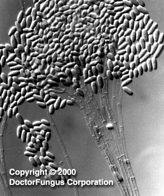
(described by Corda in 1837)
Taxonomic Classification
Kingdom: Fungi
Phylum: Ascomycota
Class: Euascamycetes
Order: Microascales
Family: Microascaceae
Genus: Graphium
Description and Natural Habitats
Graphium is a filamentous fungus found in soil and plant material. While Graphium may be isolated as an occasional contaminant, its telemorphs, Petriella, Pseudallescheria, and Ceratocystis may cause diseases. Most isolates of Graphium isolated in the clinical laboratory are synanamorphic forms of Pseudallescheria boydii or secondary forms with Scedosporium apiospermum [531, 1295].
Species
The most commonly known species of the genus Graphium are Graphium eumorphum, Graphium fructicola and Graphium putredinis.
Pathogenicity and Clinical Significance
Since one of the telemorphic forms of Graphium is included in genus Pseudallescheria, the pathologies caused by Pseudallescheria boydii in humans are relevant for the genus Graphium as well. In addition, a mixed subcutaneous infection in a captive dolphin has been found to be due to Petriella setifera, the other telemorphic state of Graphium [1814].
Notably, GR135402, a compound with antifungal activity against Candida albicans and Cryptococcus neoformans, has been isolated from a fermentation broth of Graphium putredinis. The antifungal activity of GR135402 is via inhibition of fungal protein synthesis [1193].
Macroscopic Features
Graphium colonies grow moderately rapidly and mature within 7 days. The texture is woolly to cottony. From the front, it is gray in color. From the reverse, the color is pale initially and becomes darker as the colony gets older [1295].
Microscopic Features
Graphium produces hyphae, conidiophores, synnemata, conidia, and rhizoid-like structures. Hyphae are septate and conidiophores are simple, long, and dark in color. Synemmata are bundles of erect hyphae and conidiogenous cells bearing conidia. The conidia are one-celled, oval, and colorless. They form clusters at the apex of each synnema. Rhizoid-like structures may be observed at the base of the synnema. Graphium spp. are recognized by their distinctive, erect, black synnemata, each bearing a single, terminal, ball of one-celled, hyaline conidia produced from annellides [1295].
Histopathologic Features
See our histopathology page for histopathologic features in infections due to Pseudallescheria boydii.
Compare to
Pesotum
Phialographium
Graphium must be distinguished from Pesotum, which produces sympodial conidiophores; and also from Phialographium, which produces phialides.
Laboratory Precautions
No special precautions other than general laboratory precautions are required.
Susceptibility
No data are available for Graphium spp. For susceptibility patterns of the telemorph Pseudallescheria boydii, see our N/A(L):susceptibility database.

