(Megnin) Grigorakis, 1929
Macroscopic morphology
The growth rate of the colonies is moderately rapid, reaching a diameter of 1-3 cm on Sabouraud dextrose agar incubated at 25°C for 7 days. The texture is velvety to woolly or cottony and more or less wrinkled. The color is white to gray and turns pink to buff with age. It produces a strawberry-red diffusing pigment.
Microscopic morphology
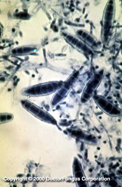
Microsporum gallinae produces septate hyphae, macroconidia and microconidia. Macroconidia and microconidia may be few or abundant. Macroconidia are clavate to cigar-shaped and 2- to 10-celled. Most commonly, 5-6 cells are observed. They are often slightly curved with fine echinulations at their apices. Microconidia are unicellular and ovoid to pyriform in shape.
Special notes
In vitro hair perforation test is negative. No special growth factor is required to grow Microsporum gallinae. It differs from Trichophyton megninii by not requiring L-histidine for growth and by producing echinulate macroconidia. Similar to Microsporum gallinae, Myxotrichum deflexum also produces a red diffusing pigment. However, this fungus does not produce conidia and has black hyphae formed after an incubation of 2-3 weeks. While Microsporum gallinae is a rare cause of tinea capitis and tinea corporis in humans, it is frequently isolated from ringworm of chickens and other fowl.