Taxonomic classification
Kingdom: Fungi
Phylum: Zygomycota
Order: Mucorales
Family: Mucoraceae
Genus: Rhizomucor

Rhizomucor miehei is often homothallic
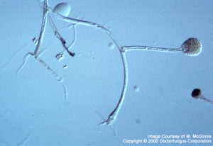
Primitive rhizoids of Rhizomucor pussilus are visible at the tips of the vegetative hyphae
Description and Natural Habitats
Rhizomucor is a cosmopolitan filamentous fungus found in soil and decaying fruit and vegetables. While Rhizomucor spp. are often isolated from fermenting and composting organic matter, they are also rare causes of serious (and often fatal) infections in humans. Rhizomucor spp. other than Rhizomucor variabilis are thermophilic in nature and can grow at temperatures as high as ~54 °C [531, 1295, 2144, 2202].
Species
The genus Rhizomucor contains three species; Rhizomucor pusillus, Rhizomucor miehei, and Rhizomucor variabilis. Rhizomucor variabilis is phylogenetically very close to Mucor hiemalis [2341]. Maximum growth temperature, biochemical assimilation profile, thiamine dependency, and the diameter of the sporangia aid in differentiation of the three Rhizomucor species from each other. Rhizomucor miehei is homothallic while Rhizomucor pusillus is homo- or heterothallic [531].
See the list of synonyms for Rhizomucor spp.
Pathogenicity and Clinical Significance
Rhizomucor spp. are among the fungi causing the group of infections referred to as zygomycosis. Although the term mucormycosis has often been used for this syndrome, zygomycosis is now the preferred term for this angio-invasive disease. Vascular invasion that causes necrosis of the infected tissue, and perineural invasion are the most frustrating features of these infections. Zygomycosis is often fatal [696, 1918].
There are few reports on human infections due to Rhizomucor spp. Cutaneous, pulmonary, rhinofacial, and disseminated zygomycosis due to Rhizomucor pusillus have so far been reported in neutropenic patients with hematological malignancies [251, 2143] and diabetes mellitus [2427]. Noteworthy, primary cutaneous infections due to Rhizomucor variabilis have been reported in otherwise healthy individuals [2493, 2494]. In contrary to the rare human infections, animal infections, particularly bovine mycotic abortion due to Rhizomucor are common [1219].
Macroscopic Features
Colonies of Rhizomucor grow very rapidly, fill the Petri dish, and mature in 4 days. The texture is typically cotton-candy like. From the front, the color of the colony is white initially and turns grey to yellowish brown in time. The reverse is white to pale. Significantly, Rhizomucor spp. other than Rhizomucor variabilis are thermophilic and yield good grow at temperatures as high as ~54 °C [531, 1295, 2144, 2202].
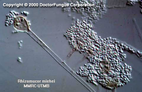
Microscopic Features
The microscopic morphology of Rhizomucor appears to be intermediate between that of Rhizopus and Mucor. Nonseptate or sparsely septate broad hyphae, rudimentary rhizoids, sporangiophores, sporangia, and sporangiospores are visualized. Rudimentary rhizoids, if exist, are few in number and are located on stolons between the sporangiophores. Sporangiophores are irregularly branched and end in sporangia at their apices. Sporangia (40-80 µm in diameter) are brown in color and round in shape. Apophysis is absent. Columellae, on the other hand, are prominent and spherical to pyriform in shape. Sporangiospores (3-4 µm in diameter) are small, unicellular, and round to ellipsoidal in shape. Zygospores, if present, are formed in the aerial hyphae. They are round to slightly compressed and dark brown to blackish brown in color [531, 1295, 2144].
Histopathologic Features
Broad, thin-walled, hyaline, often aseptate or sparsely septate hyphae are observed in the infected tissues. The contours of the hyphae are typically non-parallel and branches are irregular. Invasion of blood vessels is noteworthy [462, 763]. See also our histopathology page.
Compare to
Rhizomucor spp. must be differentiated from the other genera included in the phylum Zygomycota:
Absidia
Apophysomyces
Rhizopus
Mucor
Mortierella
Rhizomucor differs from Mucor by growing at 50-55°C and by having rhizoids and stolons; from Rhizopus by having branched sporangiophores and rhizoids not arising opposite the sporangiophores; and from Absidia by having globose sporangia and sporangiophores that are not swollen where they merge with the columellae.
The features that help in differentiation of these genera are summarized in the table below [531, 1295, 2144, 2202].
| Genus | Best growth | Sporangiophore | Apophysis | Columella | Sporangium | Rhizoid | Stylospore |
|---|---|---|---|---|---|---|---|
| Absidia | 45°C | Branched, hyaline | Conical, not prominent | Dome-shaped | Pear-shaped | +, but usually indistinct | – |
| Apophysomyces | >42°C | Usually unbranched, grayish-brown | Bell-shaped, not prominent | Usually dome-shaped, rarely elongated | Pear-shaped | + | – |
| Mortierella | 40°C | Branched, hyaline | – | – | Spherical | + | +/- |
| Mucor | <37°C | Branched or unbranched, hyaline | – | +, in varying shapes | Spherical | – | – |
| Rhizomucor | 54°C | Branched, brown | – | Spherical | Spherical | + | – |
| Rhizopus | 45°C | Unbranched and brown mostly | Not prominent | Spherical or elongated | Spherical | + | – |
Laboratory Precautions
No special precautions other than general laboratory precautions are required.
No in vitro susceptibility data are available for Rhizomucor spp.
Similar to zygomycosis due to other fungi of class zygomycetes, treatment of Rhizomucor infections consists of antifungal and surgical therapy. Amphotericin B is the most commonly used antifungal agent. Liposomal amphotericin B has also been succesfully used in some cases with zygomycosis due to Rhizomucor [251]. Early diagnosis and treatment are crucial and mortality rate is high [1918].