(described by Saccardo ex Castellani and Chalmers in 1919)
Taxonomic classification
Kingdom: Fungi
Phylum: Ascomycota
Class: Euascomycetes
Order: Microascales
Family: Microascaceae
Genus: Scedosporium
Description
Scedosporium is a filamentous fungus which occasionally causes infections in humans.
Species
The genus Scedosporium contains two species; Scedosporium apiospermum and Scedosporium prolificans. Pseudallescheria boydii is the telemorph of Scedosporium apiospermum. No sexual form (telemorph) is known for Scedosporium prolificans.
See the list of obsolete names, synonyms, and telemorphs for Scedosporium spp.
Scedosporium apiospermum
See our page on Pseudallescheria boydii for detailed information about Scedosporium apiospermum and its telemorph.
Scedosporium prolificans
Description and Natural Habitats
Scedosporium prolificans is a dematiaceous filamentous fungus isolated from soil samples.
Scedosporium prolificans can infect both immunocompetent and immunocompromised hosts. Subcutaneous infections, osteomyelitis, and arthritis are usually posttraumatic and may affect otherwise healthy individuals. Disseminated infections, on the other hand, are mostly encountered in patients who are immunosuppressed (particularly, neutropenic) due to various reasons and are often fatal [228, 908, 1090, 1405, 1826]. Scedosporium prolificans is now recognized as the most common cause of disseminated phaeohyphomycosis
[1902]. Cases with pneumonia [228], meningoencephalitis [228], and endocarditis [527] have been reported. Ocular infections (keratouveitis) [133] and colonization [1090] by Scedosporium prolificans have also been reported.
Macroscopic Features
Colonies of Scedosporium prolificans grow rapidly at 25°C and mature within 5 days. The texture is cottony and moist (yeast-like) initially which later becomes flat with fine, short, mycelial tufts. From the front, the color is light gray to black and becomes dark gray to black as the colony matures. The reverse is gray to black [1295, 2202].
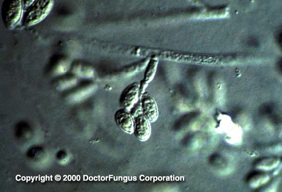
Microscopic Features
Septate hyaline hyphae, conidiogenous cells (annelides), and conidia are visualized. Annelides (conidiogenous cells) may arise directly from hyphae or are formed at the tips of the conidiophores. These annelides are flask-shaped and have a swollen base part and an elongated neck. Conidia (2-5 x 3-13 µm) are unicellular, oval-shaped, olive to brown, and have a slightly narrowed, truncated base. They are formed in clusters at the apices of the annelides. In addition, some isolates may produce round, thick-walled conidia which arise directly from the hyphae [1295, 2202].
Histopathologic Features
(Mats of) septate hyphae may be observed in the infected tissues [2061, 2148]. Scedosporium prolificans cannot be differentiated from Scedosporium apiospermum in tissue sections.
See also our histopathology page.
Compare to
Colonies of Scedosporium prolificans are darker compared to those of Scedosporium apiospermum. The inflated conidiogenous cells (annelides) and slightly wider conidia of Scedosporium prolificans, and the inability of Scedosporium prolificans to assimilate ribitol, xylitol, and L-arabinitol also help in differentiation of the two species. Besides, unlike Scedosporium apiospermum, Scedosporium prolificans does not convert to a sexual form [531, 2202]. Differentiation of these two species has been achievable also by PCR assay and hybridization probes [2389].
Laboratory Precautions
No special precautions other than general laboratory precautions are required.
Susceptibility
Scedosporium prolificans is resistant to amphotericin B [665, 687], flucytosine,
ketoconazole, miconazole,
fluconazole, and itraconazole [228, 687, 1131]. Voriconazole [495, 687], teh novel triazole Syn-2869 [1131] and caspofungin [558] also have no or very limited in vitro activity against isolates of Scedosporium prolificans.
While itraconazole or terbinafine alone has no activity against most Scedosporium prolificans isolates, the combination of these two drugs proved to be synergistic in vitro against 95% of the isolates after 48 h of incubation. In addition and importantly, antagonism was not observed in this combination study [1512].
For MICs of various antifungal drugs for Scedosporium prolificans, see our N/A(L):susceptibility database.
Due to its primary multi-resistant nature, treatment of Scedosporium prolificans infections is difficult. Amphotericin B therapy alone or in combination with flucytosine, fluconazole, or itraconazole have so far been used in cases infected with Scedosporium prolificans. However, mortality rate has been very high in most of the cases with disseminated infection [228]. Liposomal amphotericin B combined with G-CSF appeared to improve survival in an immunocompromised murine model with disseminated Scedosporium prolificans infection [1697]. Posttraumatic infections, such as arthritis in immunocompetent cases have responded to fluconazole or surgical debridement alone [1793].
Conclusively, optimal treatment of Scedosporium prolificans infections remains yet unknown and there is a great demand for novel agents with favorable activity. Importantly, the clinical outcome is closely associated with the immune status of the host, extent of the infection, and feasibility of concomitant surgical debridement.