
(described by Medlar in 1915)
Taxonomic classification
Kingdom: Fungi
Phylum: Ascomycota
Class: Euascomycetes
Order: Chaetothyriales
Family: Herpotrichiellaceae
Genus: Phialophora
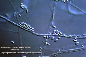
Phialophora repens, tapering phialides with parallel collarettes and curved to oval-shaped conidia
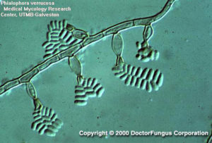
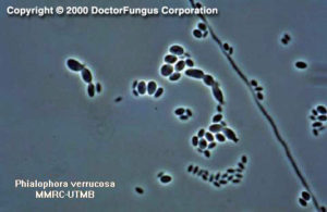
Phialophora verrucosa, dark pigmented phialides with a vase-shaped collarette
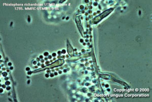
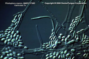
Phialophora richardsiae, phialides with saucer, flaring collarette that produces round to slightly oval conidia and on occasion some isolates will produce a second type of phialide which is similar to those of P. repens and its conidia.
Phialophora verrucosa, some isolates have a yeast like synanamorph which will produce a phialide within the yeast cell.
Phialophora parasitica, tapering, dark at the base phialides with parallel collarettes.
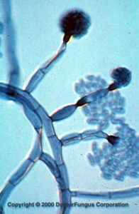
Phialophora verrucosa
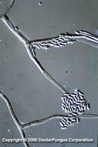
Phialophora repens
Description and Natural Habitats
Phialophora is a dematiaceous filamentous fungus that inhabits the soil, plants [2194], and decaying food. It is widely distributed in nature. Phialophora species are the causative agents of some human infections.
Species
The genus Phialophora contains 8 active species; Phialophora americana, Phialophora bubakii, Phialophora europaea, Phialophora parasitica, Phialophora reptans, Phialophora repens, Phialophora richardsiae, and Phialophora verrucosa. Phialophora europaea is a recently introduced species [532]. Morphological features, such as the shape of the collaretes, organization of the phialides, existence of chlamydospores, and biochemical features, such as the assimilation of melibiose help in differentiation of the species from each other [531].
See the list of obsolete names, synonyms, and telemorphs for Phialophora spp.
Pathogenicity and Clinical Significance
Phialophora species are among the causes of chromoblastomycosis and phaeohyphomycosis. Phialophora verrucosa is the principal causative agent of chromoblastomycosis in tropical and subtropical areas, particularly at Japan and South America. The clinical forms of phaeohyphomycosis may be diverse, including cutaneous infections, subcutaneous cysts, keratitis, endocarditis, arthritis, osteomyelitis, cerebral infection, fatal hemorrhage, and disseminated infection [602, 1388, 2053]. Phialophora europaea has been isolated from cutaneous and nail infections in North-western Europe [532].
Macroscopic Features
Colonies of Phialophora grow moderately slowly and attain a diameter of 2-3 cm following an incubation of 7 days at 25°C. The texture is wooly to velvety and may be heaped and granular in some strains. From the front, the color is initially white and later becomes dark grey-green, brown or black. From the reverse, it is iron grey to black [1295, 2144, 2202].
Microscopic Features
Septate hyphae, phialides, and conidia are observed. The hyphae (up to 5 µm wide) are branched, and hyaline to brown. Phialophora parasitica typically produces hyphae with verrucose walls.
In strains of Phialophora, conidial formation is Phialophora type. The phialides are usually flask- or bottle-shaped, pale brown to brown, and are terminally or laterally located on the hyphae. The length of the phialides may vary. Phialides of Phialophora parasitica are longer than 20 µm and spine-shaped while those of Phialophora repens are shorter than 20 µm. Phialides of Phialophora typically have clearly visible collarettes at their tips. The shape of the collarette varies from one Phialophora species to other. It is vase-shaped in Phialophora verrucosa, saucer- or vase-shaped in Phialophora richardsiae, and narrow with almost parallel contours in Phialophora repens and Phialophora parasitica. Conidia are unicellular, hyaline or brown, smooth, and round, oval or cylindrical in shape. These conidia accumulate in masses at the apices of the phialides with collarettes, giving the appearence of a vase of flowers [1295, 2144, 2202].
Histopathologic Features
Similar to the other fungi causing chromoblastomycosis, spherical or polyhedral, dark brown, thick-walled sclerotic bodies (muriform cells) are visualized in tissues infected with Phialophora species. Absence of muriform cells has been reported in cases who are immunosuppressed or debilitated [2053]. Phaeoid hyphae may also be observed.
See also our histopathology page.
Compare to
Exophiala
Fusarium
Lecythophora
Phaeoacremonium
Phialemonium
Wangiella
Phialophora differs from Exophiala by having phialides and from Wangiella by having phialides with collarettes.
Key for the differentiation of some species of Phialophora, Phialemonium, and Lecythophora
1. Phialides with saucer-shaped or flared collarettes. Phialophora richardside
1′ Phialides without saucer-shaped or flared collarettes. (2)
2. Collarettes vase-shaped, darkly pigmented; phialides flask shaped. (Phialophora verrucosa)
2′ Collarettes not vase shaped, but with parallel walls. (3)
3. Colonies first yeast-like, flat, cream-colored, turning pink to salmon (old cultures turning black in the center); phialides not separated from hyphae by a septum; conidia one-celled, often curved. (Lecythophora hoffmannii)
3.’ Colonies yeast-like (some), flat, fast growing, white, cream-colored, light gray, some with green color or light vinaceous color; tapering phialides without a basal septum, branching phialides; conidia one-celled, obovate or curved in shape. (Phialemonium spp.)
3.’ Colonies not yeast-like, gray. (4)
4. Phialides cylindrical to obclavate, elongate, hyaline to pale brown. (Phialophora parasitica)
4.’ Phialides cylindrical to lageniform, short, hyaline to pale brown. (Phialophora repens)
Laboratory Precautions
No special precautions other than general laboratory precautions are required.
Susceptibility
Very limited data are available. These data suggest that amphotericin B, itraconazole, terbinafine, and voriconazole are active in vitro against Phialophora americana, Phialophora repens, Phialophora richardsiae, and Phialophora verrucosa. The MICs of amphotericin B and itraconazole are relatively higher for Phialophora parasitica compared to other species [1490, 1492]. Voriconazole, on the other hand, is active against Phialophora parasitica as well as other Phialophora species as noted [1130]. The novel triazole, Syn-2869 is also active against Phialophora parasitica [1131]. Posaconazole shows promising activity against Phialophora species [683].
For MICs of various antifungal drugs for Phialophora, see our N/A(L):susceptibility database.